What Blood Can Tell Us About Cancer
Hemoglobin (Hb) and Cancer-Associated Anemia
A persistent drop in hemoglobin levels — especially in the absence of obvious causes like menstruation or bleeding ulcers — should raise concern. In oncology, anemia often emerges in one of three contexts:
- Chronic blood loss (e.g., colorectal cancer)
- Bone marrow infiltration (e.g., leukemia, lymphoma, metastasis)
- Chronic disease anemia (linked to inflammatory cytokines suppressing erythropoiesis)
Low hemoglobin can be the first lab abnormality in gastrointestinal or renal tumors, especially in older patients with vague fatigue.
| Parameter | Normal Range (M/F) | Concerning Trend | Potential Cancer Implication |
|---|---|---|---|
| Hemoglobin | 130–170 / 120–150 g/L | ↓ < 100 g/L | Colon, kidney, bone marrow involvement |
White Blood Cell Count (WBC) in Hematologic and Solid Tumors
Abnormalities in white blood cell count can appear as either leukocytosis or leukopenia. In acute leukemias, WBC may be dramatically elevated, often exceeding 50,000/μL. In contrast, advanced cancers or marrow failure (e.g., after metastasis) may cause profound leukopenia.
In chronic solid tumors, mild leukocytosis may reflect tumor-related inflammation or paraneoplastic leukemoid reactions.
| Parameter | Normal Range | Abnormal Trend | Possible Causes |
|---|---|---|---|
| WBC | 4.0–10.0 ×10⁹/L | ↑ > 20 ×10⁹/L | Leukemia, paraneoplastic syndrome |
| ↓ < 3 ×10⁹/L | Marrow suppression, lymphoma |
Neutrophil-to-Lymphocyte Ratio (NLR) as Prognostic Marker
Rather than looking at absolute counts, the NLR has emerged as a dynamic marker of systemic inflammation and poor prognosis. In many cancers (colorectal, lung, gastric), a high NLR (e.g., >3.5) correlates with tumor progression and poor survival. It reflects neutrophil-driven inflammation and lymphocyte-mediated immune suppression.
This ratio is especially helpful in pre-operative assessments and treatment planning.
| Parameter | Normal NLR | Elevated NLR | Clinical Interpretation |
|---|---|---|---|
| Neutrophil / Lymphocyte | ~1.5–2.5 | >3.5 | Inflammation, immune dysfunction, cancer |
Platelets (PLT) and Paraneoplastic Thrombocytosis
Platelets can both reflect and fuel cancer processes. Elevated platelet counts (thrombocytosis) are associated with tumors such as lung, ovarian, and GI cancers, often due to tumor secretion of IL-6. Inversely, low platelet counts may signal bone marrow infiltration or hematologic malignancy.
Thrombocytosis is a poor prognostic marker in many cancers and may predict metastasis.
| Parameter | Normal Range | Abnormal Level | Implication |
|---|---|---|---|
| Platelets | 150–400 ×10⁹/L | ↑ > 450 ×10⁹/L | Lung, ovarian, colon cancer |
| ↓ < 100 ×10⁹/L | Leukemia, marrow failure |
Erythrocyte Sedimentation Rate (ESR) and Cancer Inflammation
ESR is a crude but sensitive marker for chronic inflammation. Many cancers — particularly lymphomas, multiple myeloma, and solid tumors with large necrotic cores — are associated with persistently elevated ESR. Its rise is due to altered plasma proteins (especially fibrinogen), often in response to cytokines like IL-1 and TNF-alpha.
Though non-specific, ESR over 50 mm/hr, especially in the absence of infection, often prompts imaging in oncology.
| Parameter | Normal Range (M/F) | High Risk Level | Common Cancer Context |
|---|---|---|---|
| ESR | <15 / <20 mm/hr | >50 mm/hr | Myeloma, lymphoma, solid tumors |
C-Reactive Protein (CRP): The Acute-Phase Driver in Tumors
CRP rises sharply in systemic inflammation. In cancer, elevated CRP correlates with tumor burden, necrosis, and cytokine release. It is especially high in pancreatic cancer, renal carcinoma, and advanced-stage tumors. Elevated CRP at diagnosis or during therapy often correlates with worse prognosis and shorter progression-free survival.
| Parameter | Normal Range | Oncology Concern | Role in Management |
|---|---|---|---|
| CRP | <5 mg/L | >10–20 mg/L | Prognosis, treatment response |
LDH (Lactate Dehydrogenase): A Marker of Tumor Burden
LDH reflects tissue turnover and hypoxic metabolism. It is commonly elevated in lymphomas, melanomas, testicular cancer, and any cancer with aggressive growth or liver metastasis. LDH is part of the standard Ann Arbor staging for lymphomas and used in monitoring response to treatment.
Very high LDH often correlates with tumor lysis, necrosis, or hepatic involvement.
| Parameter | Normal Range | Significant Elevation | Cancer Association |
|---|---|---|---|
| LDH | <250 U/L | >500 U/L | Lymphoma, melanoma, metastases |
Ferritin: More Than Iron Storage
While ferritin is best known as an iron storage protein, in cancer it functions as an acute-phase reactant. Elevated ferritin can occur in leukemia, lymphoma, hepatocellular carcinoma, and advanced inflammation. In hemophagocytic syndromes (HLH), which can be cancer-associated, ferritin can skyrocket to thousands.
Chronically high ferritin without iron overload or liver disease should prompt oncologic evaluation.
| Parameter | Normal Range | Concerning Level | Associated Conditions |
|---|---|---|---|
| Ferritin | 20–300 ng/mL | >500–1000 ng/mL | Hematologic malignancy, HLH, liver cancer |
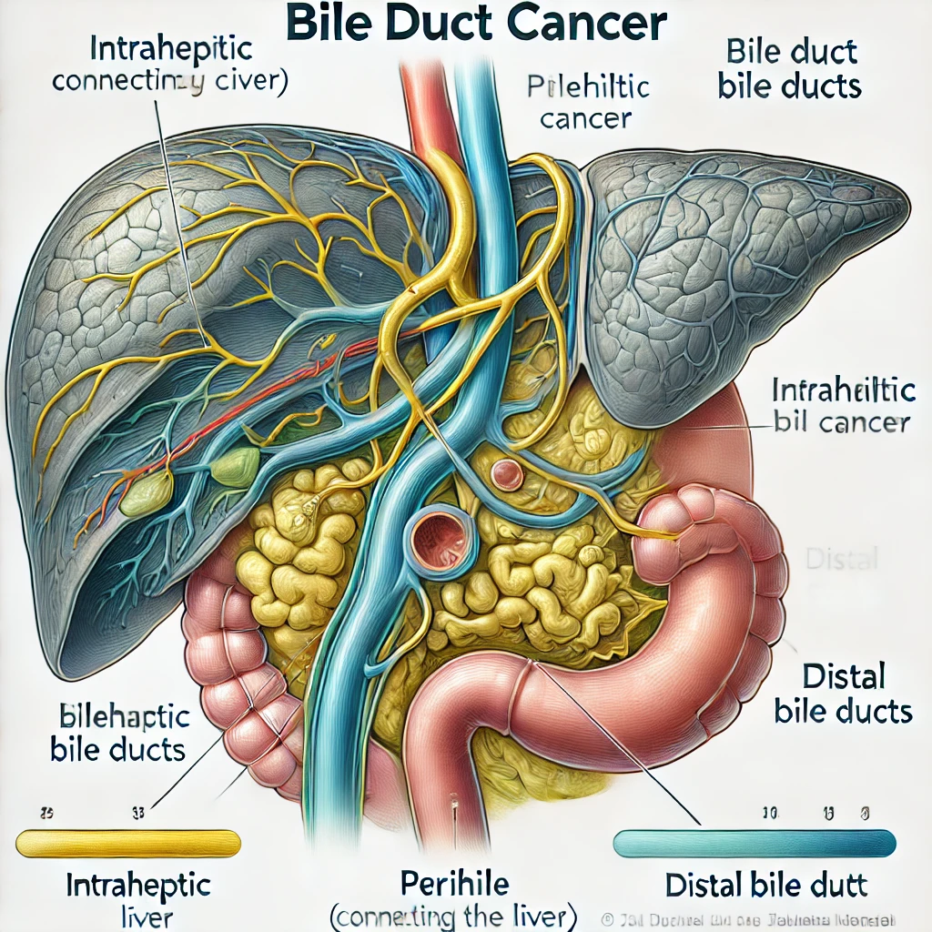
Alkaline Phosphatase (ALP): A Marker of Bone and Liver Infiltration
Alkaline phosphatase (ALP) is a key enzyme involved in bone turnover and bile duct function. While mild ALP elevation can occur in benign conditions like healing fractures or fatty liver, a persistently high ALP level — especially above twice the upper limit of normal (ULN) — may signal bone metastases or infiltration of the liver by a tumor.
In cancers like prostate, breast, lung, and multiple myeloma, ALP can be a frontline signal of skeletal involvement, often before radiologic signs appear.
In hepatobiliary tumors (e.g., cholangiocarcinoma), elevation may precede bilirubin changes, especially if bile ducts are compressed externally.
| Parameter | Normal Range (Adult) | Red Flag Values | Possible Malignancies |
|---|---|---|---|
| ALP | 30–120 U/L | >250–300 U/L | Bone mets (breast, prostate), liver tumors |
Clinical tip: If ALP is elevated alongside normal AST/ALT, consider isolated bone involvement. If ALP + GGT + Bilirubin are elevated — likely liver/biliary pathology.
AST, ALT: Transaminases in Liver-Involved Cancer
Aspartate aminotransferase (AST) and alanine aminotransferase (ALT) are intracellular enzymes released when hepatocytes are damaged. While commonly used to evaluate hepatitis and alcohol-induced liver injury, moderate elevations in AST/ALT without classic hepatitis markers may indicate:
- Liver metastases (commonly from colon, breast, or lung primaries)
- Primary liver cancers, including hepatocellular carcinoma (HCC)
- Drug-induced liver injury from chemotherapy
ALT is more liver-specific; AST is also present in muscle and cardiac tissue, so isolated AST elevation may be misleading.
| Parameter | Normal Range | Suspicious Levels | Relevance in Cancer |
|---|---|---|---|
| ALT | <35–45 U/L | >100 U/L | HCC, liver mets, chemo liver toxicity |
| AST | <35–45 U/L | >100 U/L | Less specific; interpret with ALT |
Note: In liver-dominant metastasis, transaminase elevation often coincides with fatigue, weight loss, and hepatomegaly.
Bilirubin: A Signal of Biliary Obstruction or Liver Failure
Bilirubin, a byproduct of hemoglobin breakdown, rises when bile flow is obstructed, or liver function is impaired. In oncology, elevated bilirubin may suggest:
- Obstruction by tumors (e.g., pancreatic head carcinoma compressing bile duct)
- Massive liver infiltration (e.g., in metastatic breast or colon cancer)
- Hemolysis due to cancer-related anemia or microangiopathy
Elevation of direct (conjugated) bilirubin is more common in malignancy, due to cholestasis or hepatocellular dysfunction.
| Parameter | Normal Range | Red Flags | Typical Contexts |
|---|---|---|---|
| Total Bilirubin | <1.2 mg/dL | >2.0–3.0 mg/dL | Pancreatic cancer, HCC, liver mets |
| Direct Bilirubin | <0.4 mg/dL | >1.0 mg/dL | Obstruction (cholangiocarcinoma) |
Clinical indicator: Jaundice + weight loss + elevated bilirubin = urgent imaging to rule out biliary obstruction from cancer.
Gamma-Glutamyl Transferase (GGT): Cholestasis and Tumor Pressure
GGT is an enzyme more specific to the biliary tree than ALP. While both rise in cholestasis, GGT helps differentiate liver/biliary causes from bone disease when ALP is elevated.
A high GGT with high ALP — and normal calcium/phosphate — suggests biliary obstruction or liver metastases.
| Parameter | Normal Range | High-Risk Level | Oncologic Significance |
|---|---|---|---|
| GGT | <50 U/L (men) | >150–200 U/L | Liver mets, pancreatic head tumor |
Also rises with chronic alcohol use, so context is crucial.
Tumor Markers: Context-Dependent Tools, Not Screening Tests
Tumor markers are biochemical substances secreted either by tumor cells or by the body in response to malignancy. These are not reliable for early detection, but when combined with imaging and clinical context, they provide immense diagnostic and monitoring value.
Here’s a breakdown of common markers and their use:
| Marker | Primary Use | Main Cancers | Notes |
|---|---|---|---|
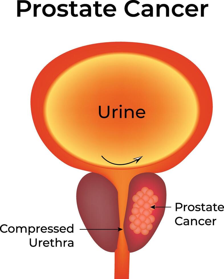
| PSA | Prostate screening and monitoring | Prostate cancer | Elevated in BPH too |
| CA-125 | Monitoring ovarian masses | Ovarian cancer | Not specific alone |
| CEA | Monitoring recurrence | Colorectal, lung, breast | ↑ in smokers |
| CA 19-9 | Pancreatic mass assessment | Pancreatic, gastric, biliary | ↑ in cholestasis too |
| AFP | Diagnosis of liver/testicular cancer | HCC, germ cell tumors | Track treatment response |
| β-hCG | Testicular, choriocarcinoma marker | Germ cell tumors, pregnancy cancers | Can be part of triple-marker panel |
| NSE | Neuroendocrine marker | Small-cell lung cancer | Less used alone |
These should never be interpreted without imaging or histology. For example, a CEA of 15 ng/mL may mean recurrence after colon cancer resection — but not in isolation.
D-dimer: Hypercoagulability as a Paraneoplastic Signal
D-dimer is a fibrin degradation product, typically used in evaluating thrombosis. However, in cancer, elevated D-dimer may be the first biochemical sign of:
- Hidden malignancy in patients with idiopathic venous thromboembolism
- Advanced-stage tumors with systemic activation of coagulation (Trousseau’s syndrome)
- High tumor burden or necrosis
| Parameter | Normal Range | Elevated Value | Associated Cancer Findings |
|---|---|---|---|
| D-dimer | <0.5 μg/mL FEU | >1.0–2.0 μg/mL | Pancreatic, gastric, lung, metastasis |
D-dimer >3.0 in a patient without thrombosis or infection should raise strong suspicion for occult malignancy.
When Blood Tells the Truth Before Imaging
Abnormalities in these blood markers are not diagnostic alone, but in combination — especially with persistent symptoms — they guide early cancer detection, staging, and even therapeutic decisions. As a clinical oncologist, I rely on these markers not just to suspect disease, but also to track the silent relapses and subtle progressions invisible on scans.
FAQ
My alkaline phosphatase (ALP) is 180 U/L. Does this mean I have bone or liver cancer?
Not necessarily. This level can be elevated due to age, medications, vitamin D deficiency, or bile duct issues. It becomes more concerning if the value rises persistently or is accompanied by symptoms like bone pain or weight loss.
My ALP rose to 270 U/L, and I have a history of breast cancer. Could it mean bone metastasis?
It could, but it’s not definitive. In patients with prior breast cancer, elevated ALP may indicate bone or liver involvement. Further imaging is usually recommended, but a single lab value without clinical signs does not confirm metastasis.
My LDH went from 210 to 265 U/L, but all scans are clear. Should I be worried?
LDH is a nonspecific marker. It can rise due to infections, inflammation, or tissue damage. Mild increases without symptoms or radiologic findings are not typically worrisome but should be monitored over time.
My hemoglobin is 9.5 g/dL and I’m male. I don’t have visible bleeding. Could it be cancer?
Yes, especially if gastrointestinal sources of blood loss have been ruled out. Low hemoglobin in the absence of clear causes should prompt evaluation for GI cancers, kidney tumors, or bone marrow involvement.
I have an ESR of 60 mm/hr. Does this always mean cancer?
No. Elevated ESR is common in infections, autoimmune diseases, and chronic inflammation. However, persistently high ESR without explanation may be a sign of lymphoma, myeloma, or other malignancies.
My CRP is consistently above 20 mg/L. Should I be tested for cancer?
Chronic CRP elevation may be related to obesity, infections, or inflammation. However, in the context of unexplained weight loss, fatigue, or other systemic symptoms, it can support suspicion of an underlying malignancy and warrants further workup.
My white blood cell count is 18,000, but I have no infection. Could it be leukemia?
A WBC count that high without infection should be investigated. While it may be due to stress or medication, leukemia or other hematologic disorders must be ruled out with further testing and possibly a bone marrow biopsy.
My platelet count is over 500,000. Is this related to cancer?
It could be. Elevated platelets (thrombocytosis) can be reactive (due to inflammation) or associated with cancers, particularly of the lung, colon, or ovaries. Persistent elevation requires evaluation.
I have very low albumin levels, but my diet is fine. Could this be from cancer?
Low albumin may indicate chronic inflammation, liver dysfunction, or cancer-related cachexia. In oncology, hypoalbuminemia is also a marker of poor prognosis in many tumor types.
Does exercise or weight loss affect CEA results?
No. Physical activity and diet do not impact CEA levels in any meaningful way.
My total bilirubin is 3.5 mg/dL and I’m yellowing. Could this be from a tumor?
Yes. Jaundice with elevated bilirubin may suggest bile duct obstruction from a pancreatic, liver, or biliary tumor. Immediate imaging is required to determine the cause.
My CA-125 is elevated but I feel fine. Should I be worried about ovarian cancer?
CA-125 can be elevated in non-cancer conditions such as endometriosis or menstruation. However, if levels are high and persistent, and especially in postmenopausal women, further pelvic imaging is necessary.
My PSA is 9.0 ng/mL but I have no symptoms. Is this prostate cancer?
An elevated PSA suggests prostate enlargement or inflammation, but cancer is possible. A prostate exam, repeat PSA, and possibly a biopsy are needed to clarify.
My CEA is slightly elevated (6.5 ng/mL) years after colon cancer surgery. Is it recurrence?
CEA is a useful marker for recurrence. A small rise may warrant repeat testing and imaging, especially if it continues to trend upward.
I had a blood clot and my doctor tested for cancer. Is that common?
Yes. Unprovoked blood clots can be an early sign of hidden cancer. This is called Trousseau’s syndrome. Some doctors order cancer screenings in these situations, especially in older patients.
My D-dimer is elevated, but I don’t have a clot. Could this be cancer?
It’s possible. D-dimer can rise in cancer, especially with metastases or active tumors. Persistent elevation without thrombosis should be evaluated further.
I’m in remission but my LDH and CRP are climbing. Is it relapse?
It might be, but not necessarily. These markers can rise due to infections or inflammation. However, in patients with a history of cancer, trends matter. Your doctor may order imaging or tumor marker testing to rule out recurrence.
My ferritin is 1200 ng/mL and I don’t take iron. Is that bad?
Yes, that level is abnormally high. Ferritin can act as an acute-phase reactant and is elevated in some cancers (lymphoma, leukemia, liver tumors). This should prompt a full diagnostic workup.