Vertigo Diagnosis and Recovery: What You Should Know
Vertigo: Definition, Types, and Risk Factors
Defining Vertigo
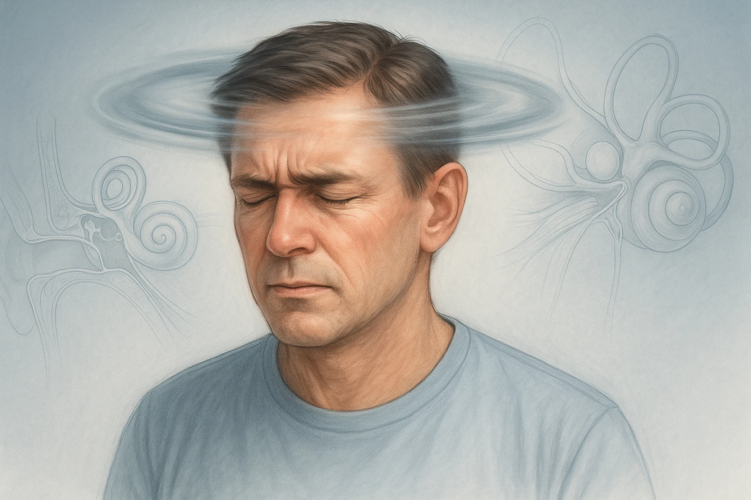
Vertigo is a false sensation of movement, most often described as a feeling that the surroundings are spinning or rotating when the body is actually still. Medically, it is recognized as a symptom rather than a disease. Vertigo differs from general dizziness or lightheadedness because it specifically involves a perceived motion, either of the patient or the environment, even in the absence of real movement.
This distinction is important because patients frequently use the term “dizziness” to describe various sensations. For clinicians, recognizing vertigo as a distinct symptom helps narrow potential causes within the vestibular or neurological systems.
Types of Vertigo
Vertigo is categorized into two main types based on its origin: peripheral and central.
| Type | Origin | Common Causes | Clinical Implications |
|---|---|---|---|
| Peripheral Vertigo | Inner ear or vestibular nerve |
|
Typically benign and self-limiting |
| Central Vertigo | Brainstem or cerebellum |
|
May indicate serious neurological disease |
Distinguishing between these two types is crucial, as peripheral causes are generally benign and self-limiting, while central causes may signal serious neurological disorders.
Prevalence and Risk Factors
Vertigo is a common clinical presentation, affecting approximately five percent of adults annually. Its incidence increases with age, reflecting age-related changes in the vestibular system and an increased likelihood of underlying conditions that disturb balance. Epidemiologic data also show that vertigo occurs more frequently in women than in men, though the exact reasons remain unclear.
- Prevalence: About 5% of adults experience vertigo each year.
- Age factor: Incidence rises with aging due to vestibular system degeneration.
- Gender difference: More common in women than men.
- Leading cause: BPPV is the most frequent form of peripheral vertigo.
Because vertigo can affect individuals across a wide age range and significantly impair daily functioning, understanding its patterns and mechanisms remains a key aspect of both diagnosis and prevention.
Causes and Mechanisms of Vertigo
Peripheral Mechanisms
Most cases of vertigo originate from peripheral disorders affecting the inner ear or the vestibular nerve, both essential for sensing motion and maintaining balance. The most frequent cause is benign paroxysmal positional vertigo (BPPV), which accounts for most peripheral vertigo cases. In BPPV, tiny calcium crystals within the inner ear become displaced, disrupting fluid movement in the semicircular canals and triggering false motion signals to the brain.
- BPPV: Displacement of calcium crystals in the inner ear causing brief spinning sensations.
- Vestibular neuritis: Inflammation of the vestibular nerve interrupting balance signals.
- Ménière’s disease: Inner ear fluid imbalance causing recurrent vertigo with hearing changes and tinnitus.
These peripheral mechanisms tend to cause sudden, intense sensations of spinning, typically worsened by head movements.
Central Mechanisms
Central vertigo arises from conditions affecting the brain’s balance centers, particularly the brainstem and cerebellum. Lesions in these regions alter the integration of sensory input from the eyes, ears, and body, producing a mismatch in perceived motion. Common central causes include:
- Stroke
- Multiple sclerosis
- Cerebellar lesions
All can impair coordination and spatial orientation. Unlike peripheral vertigo, central vertigo often presents with additional neurological signs such as imbalance, double vision, or difficulty speaking. Because central causes may signal serious disease, distinguishing them from peripheral forms is critical for safe assessment and management.
Other Contributors
Less common but important contributors to vertigo include:
- Vestibular migraine: Episodic vertigo linked to transient brain sensory dysfunction, sometimes without headache.
- Psychogenic dizziness: Perceptual imbalance related to anxiety or psychological factors.
Although these forms are less frequent than peripheral or central vertigo, recognizing them prevents misdiagnosis and ensures appropriate evaluation and reassurance. Together, these mechanisms demonstrate how vertigo arises from disruptions across both the peripheral and central balance systems.
Symptoms and Clinical Clues in Vertigo
Common Symptoms
Vertigo presents as a distinct sensation of movement or spinning, often described by patients as if the room or their body is rotating. This false sense of motion is usually accompanied by symptoms that reflect disturbances in the vestibular system.
- Dizziness or spinning sensation
- Imbalance or unsteady gait
- Nausea and vomiting
- Abnormal eye movements (nystagmus)
The intensity of symptoms can vary, with some patients experiencing brief disorientation and others reporting severe episodes that impair daily activities. Because vertigo is a symptom rather than a disease, its presentation offers important diagnostic clues. Observing whether vertigo occurs spontaneously or is triggered by movement helps clinicians identify whether the source lies in the inner ear or the central nervous system.
Time Course and Triggers
The duration and circumstances of vertigo episodes provide key diagnostic information:
- Seconds: Suggests BPPV, often provoked by head movements such as turning or lying down.
- Hours to days: Indicates vestibular neuritis or vestibular migraine, reflecting longer-lasting vestibular disturbances.
Some patients notice that vertigo worsens with changes in position, visual stimuli, or exertion, while others experience spontaneous attacks without clear triggers. Recognizing these patterns helps guide evaluation and differentiate between peripheral and central mechanisms.
Red Flags
Although vertigo is most often benign, certain features signal a possible central or serious underlying cause. Neurological symptoms such as double vision, slurred speech, weakness, numbness, or severe imbalance should prompt immediate medical attention, as they may indicate brainstem or cerebellar involvement.
- Double vision or visual disturbances
- Slurred speech
- Weakness or numbness in limbs
- Severe imbalance or difficulty walking
Vertigo with these warning signs can indicate stroke or other central nervous system pathology. Recognizing red flags early allows timely differentiation between benign peripheral causes and potentially life-threatening central disorders, ensuring safe management of patients with vertigo.
Diagnostic Approach to Vertigo
History and Physical Examination
Accurate diagnosis of vertigo begins with a detailed history and physical examination. Clinicians focus on timing, triggers, and duration of episodes, along with symptoms such as hearing loss, headache, or neurological deficits. Determining whether vertigo occurs spontaneously or after specific head movements helps narrow the differential diagnosis.
- Peripheral causes: Movement-provoked, episodic vertigo.
- Central causes: Continuous vertigo with additional neurological findings.
The physical examination includes assessment of gait, balance, and eye movements. Observation of nystagmus-rhythmic eye motion-provides valuable diagnostic clues:
- Peripheral nystagmus: Horizontal or rotational, lessens with visual fixation.
- Central nystagmus: Vertical, direction-changing, or persistent regardless of gaze.
Bedside Maneuvers
Bedside tests are essential for confirming diagnosis and guiding management. The HINTS examination-Head-Impulse, Nystagmus, Test of Skew-is a three-step assessment that distinguishes central from peripheral vertigo with high sensitivity.
| Test Component | Peripheral Vertigo | Central Vertigo |
|---|---|---|
| Head Impulse | Abnormal (catch-up saccade present) | Normal (no corrective saccade) |
| Nystagmus | Unidirectional | Direction-changing |
| Test of Skew | No vertical deviation | Skew deviation present |
The Dix-Hallpike maneuver is another key bedside test, especially for diagnosing BPPV. During this test, the patient is moved from a sitting to a lying position with the head turned, provoking characteristic vertigo and nystagmus if BPPV is present. This simple, reliable test enables both diagnosis and immediate treatment with repositioning maneuvers.
Role of Imaging
Neuroimaging is reserved for patients with atypical or central features. Magnetic resonance imaging (MRI) of the brain and posterior fossa is preferred when central pathology is suspected-such as stroke, multiple sclerosis, or tumor. Computed tomography (CT) may be used in acute settings when MRI is unavailable or contraindicated.
- Indicated: Central signs, abnormal neurological findings, or atypical presentation.
- Not routinely needed: Classic peripheral vertigo with normal neurological exam.
This selective approach ensures efficient resource use while avoiding unnecessary tests. Combining thorough clinical evaluation with targeted bedside maneuvers and imaging enables accurate, timely distinction between peripheral and central causes of vertigo.
Vertigo Treatment, Recovery, and Prevention
First-Line and Supportive Treatments
The management of vertigo depends on identifying its underlying cause. For benign paroxysmal positional vertigo (BPPV), canalith repositioning maneuvers such as the Epley or Semont procedures are considered first-line treatments. These maneuvers guide displaced calcium crystals within the inner ear back to their correct position, effectively restoring normal balance perception. Most patients experience rapid improvement after one or two sessions, though some may require repeat procedures.
- Canalith repositioning maneuvers: Epley and Semont procedures are first-line for BPPV.
- Vestibular suppressants: Short-term use (e.g., antihistamines, benzodiazepines) for acute relief, not as primary therapy.
- Central vertigo management: Requires treatment of the underlying neurological condition such as stroke or demyelinating disease.
Timely recognition and intervention are critical for improving outcomes in vertigo-related cases.
Rehabilitation and Follow-Up
Vestibular rehabilitation therapy plays a key role in recovery, especially for patients with persistent imbalance or those recovering from central causes. This exercise-based program helps the brain recalibrate balance and visual cues, improving coordination and confidence in daily movement.
- Goals of rehabilitation: Recalibrate balance and visual processing.
- Approach: Gradual re-exposure to motion and balance exercises to restore stability.
- Follow-up: Repeat maneuvers or therapy sessions if symptoms recur.
Patient reassurance is also essential, as vertigo can be distressing and often misunderstood. Educating patients about the benign nature of most peripheral causes and providing guidance on expected recovery times can significantly reduce anxiety. Regular follow-up enables clinicians to monitor progress and repeat repositioning maneuvers if symptoms reappear.
Prognosis and Prevention
Most cases of peripheral vertigo resolve with appropriate maneuvers or rehabilitation, often within days to weeks. Central vertigo outcomes vary depending on the underlying neurological disorder, but early diagnosis and rehabilitation can support functional recovery.
- Prognosis: Peripheral vertigo generally resolves with treatment; central causes depend on underlying pathology.
- Recurrence: Common in BPPV but manageable with repeat repositioning and vestibular exercises.
- Prevention: Maintain regular physical activity, avoid prolonged bed rest, and continue vestibular exercises to prevent recurrence.
Patients who understand their condition and actively participate in rehabilitation tend to recover more quickly and experience fewer recurrences. A combination of reassurance, targeted therapy, and consistent follow-up forms the foundation of effective vertigo management.
Key Clinical Insights for Vertigo Learners
Core Learning Points
Vertigo assessment begins with a detailed history and physical examination. Understanding the timing, triggers, and associated symptoms helps narrow the differential diagnosis between peripheral and central causes. Peripheral vertigo, such as benign paroxysmal positional vertigo (BPPV), often presents with brief, position-triggered episodes, while central vertigo tends to be continuous and accompanied by neurological findings. Recognizing these distinctions early prevents unnecessary testing and guides efficient care.
- Peripheral vertigo: Brief, movement-triggered episodes (e.g., BPPV).
- Central vertigo: Continuous symptoms with neurological signs.
- Best practice: Begin with bedside assessment before imaging.
Common diagnostic errors include relying solely on imaging or misinterpreting dizziness as vertigo without confirming a true sense of motion. A methodical approach emphasizing bedside evaluation over early imaging ensures more accurate and cost-effective diagnosis.
Practical Clinical Application
The HINTS examination-comprising the Head Impulse test, Nystagmus evaluation, and Test of Skew-is a high-sensitivity bedside tool for differentiating peripheral from central vertigo. When performed correctly, it can outperform early MRI in detecting posterior circulation strokes. In patients with features consistent with BPPV, the Dix-Hallpike maneuver can confirm the diagnosis, and canalith repositioning techniques such as the Epley or Semont maneuvers can provide immediate relief.
| Bedside Test | Purpose | Interpretation |
|---|---|---|
| HINTS | Distinguish central vs peripheral vertigo | Central findings suggest stroke or cerebellar lesion |
| Dix-Hallpike | Identify BPPV of posterior canal | Positive if vertigo and nystagmus are triggered |
| Epley / Semont | Treat BPPV by repositioning otoliths | Usually resolves symptoms within one or two sessions |
Neuroimaging, typically MRI, should be reserved for patients with atypical symptoms, persistent imbalance, or neurological deficits suggestive of central involvement. Recognizing when to escalate care-such as in cases of double vision, weakness, or new-onset gait disturbance-is an essential part of safe and effective vertigo management.
Questions to Ask During Training
For learners developing vestibular assessment skills, reflective questioning can deepen clinical understanding:
- What historical clues distinguish vertigo from general dizziness or presyncope?
- How can bedside tests like HINTS and Dix-Hallpike help avoid unnecessary imaging?
- When is it appropriate to refer a patient for specialist evaluation or neuroimaging?
- What strategies improve patient reassurance and adherence to repositioning exercises?
Engaging with these questions during supervised practice helps medical trainees integrate vestibular physiology, examination technique, and clinical reasoning into safe and confident patient care.
Frequently Asked Questions About Vertigo
- Is vertigo the same as dizziness?
- No. Vertigo involves a false sense of movement or spinning, while dizziness is a broader term that may describe lightheadedness or imbalance without motion.
- What usually causes vertigo?
- Most cases come from inner ear conditions such as benign paroxysmal positional vertigo (BPPV), vestibular neuritis, or Ménière’s disease. Less commonly, it results from central brain disorders like stroke.
- How long can vertigo episodes last?
- The duration depends on the cause. BPPV episodes last seconds, vestibular neuritis may persist for days, and central vertigo can be more continuous.
- Are there warning signs that vertigo might be serious?
- Yes. Symptoms like double vision, slurred speech, limb weakness, or severe imbalance may indicate a neurological problem requiring urgent evaluation.
- How is vertigo diagnosed by doctors?
- Diagnosis relies on a detailed medical history, physical examination, and bedside tests such as the HINTS or Dix-Hallpike maneuvers. Imaging is reserved for atypical or central presentations.
- What treatments are effective for vertigo?
- For BPPV, canalith repositioning maneuvers like the Epley or Semont procedures are first-line. Vestibular rehabilitation and short-term symptom relief medications may also help recovery.
- Can vertigo come back after treatment?
- Yes, recurrence is common in BPPV but manageable. Repeating repositioning maneuvers and maintaining balance exercises can reduce future episodes.
- Does age increase the risk of vertigo?
- It does. Aging affects the vestibular system and increases the likelihood of conditions that disturb balance, making vertigo more frequent in older adults.
- Is vertigo linked to stress or anxiety?
- Stress does not directly cause vertigo but can worsen perception of dizziness. Anxiety-related or psychogenic dizziness can mimic vertigo sensations.
- Can exercise help prevent vertigo?
- Regular movement and vestibular exercises can support balance and reduce recurrence. Avoiding prolonged bed rest after an episode also promotes faster recovery.


