Dry eyes: causes, symptoms, and treatment options
Dry eye disease: definition, who is affected, and clinical presentation
Dry eye disease (DED) (dry eyes) is a multifactorial condition characterized by bothersome ocular symptoms alongside tear film instability and loss of homeostasis. In clinical settings, this framework helps explain why patients report discomfort and visual fluctuation during routine tasks. At its core, DED reflects interacting processes: tear film hyperosmolarity, ocular surface inflammation, and neurosensory abnormalities. These mechanisms shape both the pattern of symptoms and the clinical findings observed at the slit lamp. Understanding the major subtypes-aqueous-deficient and evaporative-clarifies etiology and guides management, while awareness of who is most affected and which risks are relevant supports targeted history taking. Common symptoms such as dryness, burning, foreign body sensation, blurred or fluctuating vision, and photophobia should prompt structured assessment.
Modern Definition and Mechanisms
DED is defined by symptoms in the presence of tear film instability and loss of homeostatic control. This instability can disrupt the smooth optical surface required for consistent vision, aligning with patient-reported fluctuations.
Mechanistically, hyperosmolarity and ocular surface inflammation are central, with neurosensory abnormalities also contributing. These elements interact to perpetuate discomfort and visual disturbance, offering a practical lens for interpreting both presentation and response to therapy.
- tear film hyperosmolarity
- ocular surface inflammation
- neurosensory abnormalities
Subtypes and Mixed Presentations
- Aqueous-deficient dry eye arises when lacrimal secretion is reduced, as in conditions such as Sjögren syndrome. The resulting deficiency in the aqueous layer compromises tear quantity and stability.
- Evaporative dry eye most commonly reflects meibomian gland dysfunction, which increases tear evaporation by altering the lipid layer. Recognizing whether deficiency of production or excess evaporation predominates helps prioritize initial interventions.
Burden, Risks, and Symptoms
DED becomes more prevalent with advancing age and is observed more often in women. Risk factors relevant to routine clinical screening include meibomian gland dysfunction, autoimmune disease, contact lens wear, environmental stressors, and medications that reduce tear production.
- meibomian gland dysfunction
- autoimmune disease
- contact lens wear
- environmental stressors
- medications that reduce tear production
Patients frequently report dryness, burning, a foreign body sensation, fluctuating or blurred vision, and sensitivity to light (photophobia). Taken together with the mechanistic framework above, this symptom profile supports early identification and appropriate direction of diagnostic testing and management.
- dryness
- burning
- a foreign body sensation
- fluctuating or blurred vision
- sensitivity to light (photophobia)
Diagnosing dry eye disease: structured assessment and objective testing
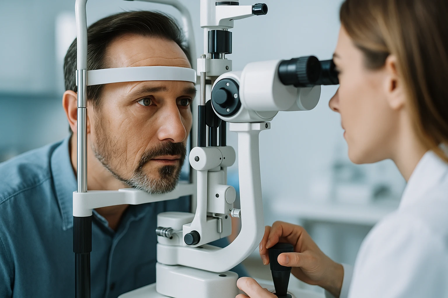
A practical diagnostic approach to dry eye disease (dry eyes) combines standardized symptom measurement with slit-lamp examination and targeted objective tests. The goal is to document how patients feel and what is observed at the ocular surface, then align these findings to characterize the disorder with clinical precision. Because symptoms may not correlate linearly with objective signs, a consistent framework helps avoid under- or overestimating disease severity and supports clear communication across visits and providers.
- Standardized symptom measurement
- Slit-lamp examination
- Targeted objective tests
Symptom Quantification
Validated instruments such as the Ocular Surface Disease Index (OSDI) or the Dry Eye Questionnaire-5 (DEQ-5) provide a repeatable baseline for symptom burden and allow comparison over time. Using these tools alongside slit-lamp examination anchors the history in measurable data, capturing the frequency and impact of dryness, visual fluctuation, and related discomfort in a way that complements clinical observation.
- Ocular Surface Disease Index (OSDI)
- Dry Eye Questionnaire-5 (DEQ-5)
Structured symptom scoring also facilitates goal setting for follow-up. When recorded consistently, changes in scores can be interpreted with the clinical examination to judge whether observed ocular surface findings align with the patient’s experience.
Core Objective Tests
Tear film break-up time (TBUT) evaluates tear film stability and helps determine whether instability contributes to visual fluctuation and discomfort. Corneal and conjunctival staining highlight areas of epithelial compromise, providing a map of surface involvement that can be compared across visits.
| Test | Primary role |
|---|---|
| Tear film break-up time (TBUT) | Evaluates tear film stability |
| Corneal and conjunctival staining | Highlights areas of epithelial compromise |
| Schirmer testing | Assesses tear production |
Schirmer testing assesses tear production and is helpful when reduced aqueous secretion is suspected. Selection and timing of these tests are informed by the initial history and slit-lamp examination, ensuring that measurements address the most relevant aspects of tear quantity and surface integrity for the patient in front of the clinician.
Integrating Signs and Symptoms
Because symptoms may not correlate linearly with objective signs, interpretation requires synthesis rather than reliance on any single measure. Documenting both patient-reported outcomes and examination findings allows clinicians to recognize presentations where symptoms are prominent despite limited staining, or where surface changes are evident despite lower symptom scores.
- Symptoms are prominent despite limited staining
- Surface changes are evident despite lower symptom scores
This integration supports subtype-directed thinking: for example, instability reflected by TBUT and surface staining can be considered alongside symptom scores and the slit-lamp examination to clarify whether evaporative features or aqueous deficiency are more likely. Consistent recording of symptom instruments and objective tests creates a clear longitudinal picture that informs subsequent clinical decisions.
Foundations of care: nonpharmacologic strategies and medical therapy
Initial management of dry eye disease (dry eyes) emphasizes simple measures that stabilize the tear film and reduce symptom triggers. Practical steps include environmental modification, planned screen breaks, and the routine use of preservative-free lubricants as needed. For patients with signs of meibomian gland dysfunction, warm compresses and lid hygiene support the lipid layer and reduce evaporative loss. When symptoms persist despite these measures, escalation to anti-inflammatory therapy may be appropriate. Throughout chronic care, minimizing preservative exposure helps protect the ocular surface.
- Environmental modification and planned screen breaks
- Preservative-free lubricants as needed
- Warm compresses and lid hygiene for meibomian gland dysfunction
- Escalation to anti-inflammatory therapy when symptoms persist
- Ongoing preservative minimization in chronic care
Environment, Behavior, and Lid Care
Modifying the environment and daily routines can lessen symptom burden and complement medical therapy. Screen breaks interrupt prolonged visual tasks that are associated with reduced blink frequency, while attention to airflow and humidity can limit evaporative stress. Incorporating these adjustments early gives patients a clear structure for self-management alongside clinical follow-up.
- Screen breaks during prolonged visual tasks
- Attention to airflow and humidity
- Lid hygiene and warm compresses are recommended, particularly when meibomian gland dysfunction is present
- Regular heat and gentle cleansing can support meibomian gland function and stabilize the tear film’s lipid component
Lid hygiene and warm compresses are recommended, particularly when meibomian gland dysfunction is present. Regular heat and gentle cleansing can support meibomian gland function and stabilize the tear film’s lipid component. These measures integrate well with other first-line steps and can be continued as part of long-term maintenance.
Lubricant Strategy
Preservative-free artificial tears are an appropriate baseline option and can be used as needed to relieve symptoms and support surface integrity. Selecting preservative-free formulations is especially important in patients who require frequent dosing or long-term use, because chronic exposure to preservatives can worsen ocular surface disease.
- Preservative-free artificial tears as baseline therapy
- Chronic exposure to preservatives can worsen ocular surface disease
- A trial-and-assess approach helps align lubricant use with symptom patterns observed over time
- Adjust frequency or formulation based on response, prioritizing preservative minimization for chronic therapy
A trial-and-assess approach helps align lubricant use with symptom patterns observed over time. Clinicians can document baseline symptom scores and adjust frequency or formulation based on response, always prioritizing preservative minimization for chronic therapy.
Escalation to Anti-inflammatory Therapy
When symptoms and signs remain active despite consistent use of preservative-free lubricants and basic measures, anti-inflammatory therapy can be considered. Topical cyclosporine or lifitegrast are options for inflammatory disease not controlled with lubricants, reflecting a stepwise approach from foundational care to targeted pharmacologic treatment.
- Topical cyclosporine or lifitegrast for inflammatory disease not controlled with lubricants
- Short courses of topical corticosteroids may improve symptoms and signs but require monitoring due to intraocular pressure risk
- If used, they should be time-limited and accompanied by follow-up to assess clinical response and safety
- Maintaining preservative minimization across the regimen remains an important principle of chronic ocular surface care
Short courses of topical corticosteroids may improve symptoms and signs but require monitoring due to intraocular pressure risk. If used, they should be time-limited and accompanied by follow-up to assess both clinical response and safety. As therapy intensifies, maintaining preservative minimization across the regimen remains an important principle of chronic ocular surface care.
Procedures, safety, and referral pathways
Procedural options in dry eye care (dry eyes) focus on conserving tears and addressing meibomian gland dysfunction (MGD), while ongoing safety monitoring and timely referral maintain patient-centered outcomes. Selection should be individualized by severity, subtype, and comorbid conditions, with expectations aligned to the current evidence base. Escalation decisions are grounded in response to stepwise therapy and the presence of red flags or suspected systemic associations.
Tear Conservation and MGD Procedures
Punctal occlusion may be used for tear conservation in selected cases. By reducing outflow, this approach aims to increase tear residence time when symptoms and signs suggest inadequate retention of tears despite foundational measures. Candidacy should be considered within a structured plan that accounts for patient subtype and overall disease burden.
For MGD-directed care, thermal pulsation and other device-based therapies have suggestive but not definitive evidence of benefit. These options can be discussed when clinical examination indicates evaporative stress linked to meibomian gland dysfunction, with counseling that current evidence supports potential improvement but remains inconclusive. When used, they should be integrated into a broader regimen that also includes ongoing nonpharmacologic measures.
| Procedure | Primary intent | Evidence posture |
|---|---|---|
| Punctal occlusion | Tear conservation in selected cases | Used within a structured plan |
| Thermal pulsation and device-based therapies | MGD-directed care | Suggestive but not definitive benefit |
Safety and Monitoring Essentials
Topical corticosteroids can play a limited adjunctive role; however, they carry risks such as intraocular pressure elevation and should be time-limited with follow-up. Monitoring should confirm that symptom changes align with examination findings and that pressure-related adverse effects are not emerging. Preserving a cautious, planned duration helps maintain a favorable risk-benefit balance.
Across chronic management, routine review of treatment exposure supports safety. As therapy evolves, documenting symptom scores alongside examination findings facilitates early recognition of discordance and guides whether procedural or medical adjustments are warranted, always maintaining a focus on preservative minimization within the overall regimen.
- Time-limited use of topical corticosteroids with follow-up
- Confirm alignment of symptom changes with examination findings
- Routine review of treatment exposure across chronic management
- Maintain focus on preservative minimization within the regimen
When to Escalate or Refer
Referral or escalation is indicated when symptoms persist despite stepwise therapy or when red flags or systemic associations are suspected. This includes scenarios where patient-reported burden remains high even as objective findings show limited change, or the reverse, and when the pattern of disease suggests a need for specialized evaluation.
Care coordination benefits from clear thresholds and individualized planning. Management should be tailored by severity, subtype, and comorbid conditions, ensuring that procedural choices, safety monitoring, and referral timing are aligned with the patient’s clinical trajectory and goals of care.
- Symptoms persist despite stepwise therapy
- Red flags or systemic associations are suspected
- High symptom burden with limited objective change, or the reverse
- Need for specialized evaluation based on disease pattern
Dry eyes: frequently asked questions
My eyes feel gritty, but tests were “normal.” Is that still dry eye?
Yes. Symptoms and exam findings don’t always match in dry eye. Clinicians combine symptom scores with slit-lamp examination and targeted tests to build the full picture.
Do contact lenses make dry eyes worse?
They can. Contact lens wear is a known risk factor because it can destabilize the tear film and increase dryness or irritation in some people.
Why does my vision fluctuate during reading or screen time?
Reduced blinking during prolonged visual tasks destabilizes the tear film. Planned screen breaks and attention to airflow and humidity can lessen these fluctuations.
What’s the difference between evaporative dry eye and aqueous-deficient dry eye?
Evaporative dry eye is commonly linked to meibomian gland dysfunction, which increases tear evaporation. Aqueous-deficient dry eye stems from reduced lacrimal secretion, lowering tear quantity.
Why are preservative-free lubricants emphasized?
Chronic exposure to preservatives can worsen the ocular surface. Preservative-free artificial tears are preferred for frequent or long-term use.
When are prescription anti-inflammatory drops considered?
They’re used when symptoms and signs persist despite preservative-free lubricants and basic measures. Options include topical cyclosporine or lifitegrast for inflammatory dry eye.
Are steroid eye drops safe for dry eyes?
Short courses can improve symptoms and signs, but they carry risks such as increased intraocular pressure. Use is time-limited with follow-up to monitor safety.
What procedures might help if drops aren’t enough?
Punctal occlusion conserves tears in selected cases by reducing outflow. Thermal pulsation and other device-based therapies target meibomian gland dysfunction, with suggestive but not definitive evidence of benefit.
When should I see a specialist for dry eyes?
Referral is appropriate when symptoms persist despite stepwise care, when red flags are present, or when systemic associations are suspected. Management is individualized by severity, subtype, and comorbid conditions.