Does Tattoo Removal Cause Cancer? Discover the Truth Behind Tattoo Removal Safety
- What Happens to Tattoo Ink in the Skin
- Types of Tattoo Removal and Their Chemical Impact
- Tattoo Ink Composition and Cancer Concerns
- Laser-Induced Chemical Reactions in Tattoo Removal (таблица)
- Regulatory Oversight and Lack of Standardization in Tattoo Inks
- Epidemiological Evidence: What Long-Term Studies Say
- How the Immune System Responds to Laser-Shattered Pigment
- Risk Differentiation by Tattoo Location, Size, and Ink Depth
- Chemical Reactions During Laser Tattoo Removal
- Understanding the Role of Ink Carriers and Additives
- Insights from Animal Studies on Tattoo-Related Carcinogenicity
- Comparative Overview: Tattoo Removal vs. Other Cosmetic Procedures
- The Role of Chronic Inflammation in Skin Health Post-Tattoo Removal
- Differences Between Laser Technologies and Their Risk Profiles
- Preventative Strategies to Minimize Long-Term Health Risks
- Research Gaps and Future Directions in Tattoo Ink Safety
- FAQ
What Happens to Tattoo Ink in the Skin
Tattoo ink sits in the dermis, the skin’s second layer, where immune cells surround and trap pigment particles. These particles remain visible through the transparent outer skin, giving tattoos their permanence. Ink composition varies widely, often including organic and inorganic compounds, heavy metals, and sometimes even plastic-based pigments. Over time, immune cells slowly break down and transport tiny pigment fragments to the lymph nodes, which can discolor them.
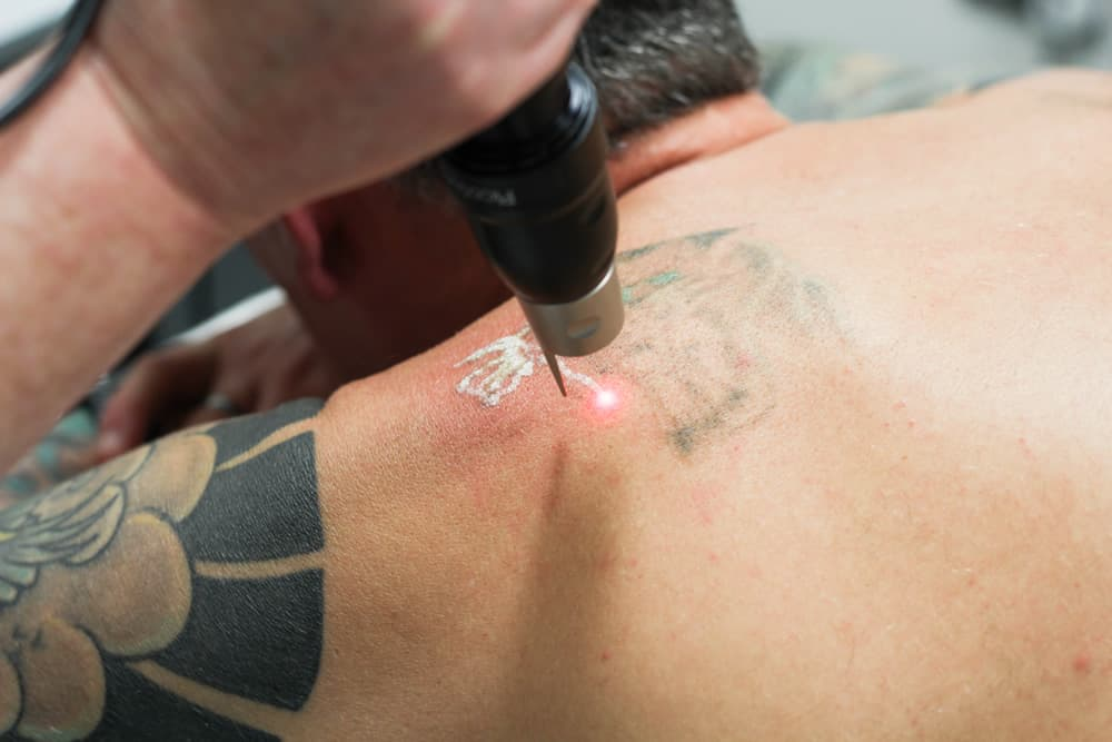
During tattoo removal, high-energy laser pulses—most often Q-switched or picosecond lasers—are used to break up the ink particles into smaller fragments. The body’s lymphatic system then gradually clears them, much like it would with debris from minor skin damage. This raises questions about what exactly is being absorbed and where it goes.
Researchers have identified that certain pigments, particularly azo dyes, can decompose into aromatic amines under laser irradiation. Some of these byproducts have been shown to be potentially mutagenic in laboratory conditions. However, the clinical significance of this transformation remains debated, and large-scale studies linking it to cancer in humans are lacking.
Types of Tattoo Removal and Their Chemical Impact
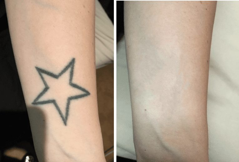
The most common method of removal is laser-based. Q-switched lasers work by emitting short pulses of light that fragment ink via photomechanical effects. Picosecond lasers are newer and deliver even shorter bursts, often considered more efficient. Both systems target pigments while sparing surrounding tissue.
When ink is shattered, its fragments are small enough to be absorbed and processed by macrophages. But this fragmentation process, especially in colored inks, may generate harmful byproducts. For instance, red, yellow, and green pigments often contain metals such as cadmium, mercury, and chromium. Laser exposure can oxidize these elements, producing compounds that, in isolated studies, exhibit carcinogenic properties.
Non-laser methods such as dermabrasion or surgical excision carry different risks. While they avoid chemical reactions caused by heat and light, they are more invasive and increase infection risk, which itself can lead to chronic inflammation—a known cancer-promoting factor.
In all methods, the level of systemic exposure to ink degradation products is extremely low. Current dermatological consensus holds that no direct link to cancer has been established, but that continued monitoring is warranted as ink compositions evolve.
Tattoo Ink Composition and Cancer Concerns
Ink formulas are not uniformly regulated. In the United States, the FDA does not actively review or approve tattoo inks before they enter the market. This leaves manufacturers to self-regulate, resulting in highly variable formulations. Some include known carcinogens like benzo(a)pyrene or phthalates, although usually at low concentrations.
Black ink, which is the most commonly used, often contains carbon black—a material that, when inhaled in industrial settings, is considered a possible human carcinogen. However, subdermal exposure (i.e., through tattooing) is very different from inhalation, and no equivalent risk level has been firmly identified for skin exposure.
[ссылка здесь: в тему органических рисков можно встроить материал про паразитов и рак груди — do parasites cause breast cancer]
It’s also worth noting that tattoo pigment particles are sometimes found in nearby lymph nodes, raising concerns about long-term immune modulation. While this isn’t directly cancerous, chronic immune activation and pigment accumulation are still being studied for their systemic effects.

Laser-Induced Chemical Reactions in Tattoo Removal
| Ink Color | Common Compounds | Possible Byproducts After Laser | Cancer Risk Classification |
| Black | Carbon black | Polycyclic aromatic hydrocarbons (PAHs) | Group 2B (possible human carcinogen) |
| Red | Cinnabar, Azo dyes | Mercury compounds, aromatic amines | Insufficient human data |
| Yellow | Cadmium sulfide | Cadmium oxides, free radicals | Known human carcinogen (inhalation) |
| Green | Chromium oxide | Chromium(VI) compounds | Group 1 (known carcinogen) |
| Blue | Cobalt aluminate | Cobalt oxides | Group 2B (possible human carcinogen) |
These reactions depend on laser wavelength, ink depth, pigment type, and even patient skin tone. However, the quantities of released byproducts are so small that cancer risk remains theoretical and unproven in clinical settings.
Regulatory Oversight and Lack of Standardization in Tattoo Inks
One of the critical concerns in evaluating cancer risks from tattoo removal is the inconsistent regulation of tattoo ink ingredients. In the United States, the Food and Drug Administration (FDA) classifies tattoo inks as cosmetics but does not actively approve or review them before use. As a result, many pigments are sourced from industrial paints or textile dyes, with little transparency about long-term biological impact.
In Europe, the EU’s REACH regulation has introduced stricter controls, banning several pigments and requiring manufacturers to disclose components. Still, the enforcement varies across countries, and tattoo parlors may use leftover stock or imported materials. This inconsistency leads to varying exposure levels of harmful substances across patients, complicating any attempt to study long-term cancer correlations.
The uncertainty lies not just in the ingredients but also in the chemical changes those ingredients undergo during laser removal. Without standardization, risk prediction becomes complex, and what’s safe in one case might not be in another. This is a growing concern as tattoo popularity rises globally, especially among younger demographics who may undergo removals decades later.
Epidemiological Evidence: What Long-Term Studies Say
To date, no large-scale epidemiological study has definitively linked laser tattoo removal to increased cancer risk. While certain case studies and laboratory models have suggested the potential for mutagenic byproducts, real-world data remains inconclusive. In studies involving thousands of tattooed individuals, cancer incidence did not appear significantly elevated compared to non-tattooed populations when controlling for variables such as smoking or UV exposure.
A 2016 review published in Archives of Dermatological Research analyzed over 300 cases of laser removal and found no statistically significant cancer development post-treatment. However, researchers emphasized the need for long-term follow-up, as some cancers may take decades to manifest. Moreover, the review identified several methodological limitations in existing research, including small sample sizes, short observation periods, and lack of control for confounding factors.
This aligns with the broader challenge of tracing chronic diseases to low-level environmental exposures. It’s difficult to isolate tattoo removal as a single variable when individuals may have occupational exposures, poor diet, or genetic predispositions that affect cancer development. The available data is therefore best interpreted as reassuring, but not conclusive.

How the Immune System Responds to Laser-Shattered Pigment
When tattoo ink is disrupted by laser energy, the resulting particles are too large to exit the skin on their own. Instead, immune cells such as macrophages engulf and transport them to lymph nodes and other filtration organs. This process is part of the body’s natural waste-management system, but it raises two key concerns: the accumulation of foreign particles and prolonged immune activation.
Chronic immune stimulation is one known factor that can contribute to carcinogenesis. In theory, if pigment residues trigger long-term inflammatory responses or disrupt immune signaling, this could influence the development of skin-related or lymphatic cancers. Yet, in practice, such cases are exceedingly rare, and most immune responses to laser removal resolve within weeks.
Histological studies have shown that pigment-loaded macrophages can remain in the dermis or lymph nodes for years without causing pathology. Nonetheless, patients with compromised immune systems or autoimmune conditions should approach laser removal with caution, as their immune surveillance may be less capable of clearing debris safely.
In sum, the immune system’s interaction with ink fragments is complex but does not, in healthy individuals, appear to increase cancer susceptibility under normal conditions.
Risk Differentiation by Tattoo Location, Size, and Ink Depth
Tattoo characteristics play a large role in determining how the body responds to removal and whether there is a theoretical risk of carcinogenic exposure. Larger tattoos involve more pigment volume, increasing the burden on the lymphatic system when fragments are cleared. Deeper ink placement, common in professional tattoos, requires more intense laser energy to reach, potentially generating more reactive breakdown products.
Visible areas like the forearm or back receive more UV exposure, which can compound the stress on skin cells after laser treatment. The face and neck have thinner skin and more delicate vasculature, making them more sensitive to inflammatory reactions post-removal.
Moreover, multicolored tattoos pose higher chemical risk due to the variety of pigments used. Blue and green inks, for instance, often contain copper-based and chromium-based compounds, respectively. When fragmented by laser, these can release compounds whose safety has not been evaluated in subdermal use.
However, individual risk remains highly variable. The ink’s original formula, the laser type and wavelength, skin tone, and immune status all contribute to how the body processes tattoo removal. Therefore, while location and size inform technical complexity, they do not, on their own, predict cancer risk conclusively.
Chemical Reactions During Laser Tattoo Removal
Laser removal works by delivering short, high-intensity pulses of light that break tattoo pigments into smaller fragments. This process is photothermal and photomechanical, meaning it generates localized heat and shockwaves within the skin. When these forces interact with complex organic and inorganic compounds found in tattoo ink, they initiate chemical reactions that are not always benign.
Recent spectrometry studies have shown that lasers can trigger the formation of new chemical species, some of which are potentially cytotoxic or mutagenic. For example, black ink often contains polycyclic aromatic hydrocarbons (PAHs), which under laser impact may degrade into benzene derivatives — substances classified as human carcinogens. Similarly, the breakdown of azo pigments (common in red, orange, and yellow inks) may release aromatic amines, another category of cancer-related compounds.
While these reactions occur in minuscule concentrations and are quickly diluted or removed by biological systems, their presence raises valid concerns for repeated exposure or poorly metabolized byproducts. Hence, the biochemical aftermath of tattoo removal is an important factor in long-term risk assessment, particularly in individuals undergoing multiple sessions or targeting large coverage areas.
Understanding the Role of Ink Carriers and Additives
Tattoo ink is not composed solely of pigment. It also includes carriers — solvents that help disperse and stabilize the pigment particles. These carriers may include glycerin, ethanol, witch hazel, propylene glycol, and water. While often assumed to be inert, some of these ingredients can react under laser energy or interact with pigments to form unstable intermediates.
One under-researched area involves the role of nanoparticles in modern inks. Some black inks use carbon black in nano-sized particles to achieve better diffusion and color saturation. These nanoparticles may penetrate deeper skin layers or behave unpredictably under laser exposure. Studies have raised concerns that nanoparticle migration through the lymphatic system might increase systemic distribution, although current evidence does not confirm a cancer risk from this process.
Carcinogenicity is not limited to pigments alone — it’s the combination of pigments, carriers, stabilizers, and their transformations under laser energy that creates a full toxicological profile. Unfortunately, labeling of ink components is not mandatory in many countries, leaving both practitioners and patients unaware of the exact substances involved in their tattoos or their breakdown.
Insights from Animal Studies on Tattoo-Related Carcinogenicity
Due to ethical limitations, many cancer-related hypotheses involving tattoo ink and laser treatment are first explored in animal models. Rodent studies, particularly in mice and rats, provide useful — though imperfect — analogs for human responses to pigment absorption and clearance.
In several long-term studies, rats injected subcutaneously with commonly used tattoo pigments exhibited low-grade chronic inflammation and pigment migration to lymph nodes. However, there were no consistent signs of tumor formation in the injection sites. Only under certain experimental conditions — such as simultaneous exposure to known carcinogens or immune-suppressing agents — did tumor development occur.
When lasers were applied to pigmented skin in animal models, the formation of heat-degraded byproducts was measurable but transient. Inflammatory markers increased temporarily, yet returned to baseline within days. These results support the conclusion that tattoo removal alone is unlikely to initiate cancer, although it could contribute to risk in a body already under carcinogenic stress.
Importantly, animal models lack the complexity of human immune modulation and lifestyle variables. Their predictive power is therefore limited, but they remain essential for identifying potential mechanisms of harm. This idea of multiple factors is well complemented by the article What is PTP1B in breast cancer?, which explains the role of biochemical pathways in oncogenesis
Comparative Overview: Tattoo Removal vs. Other Cosmetic Procedures
| Procedure Type | Tissue Impact | Chemical Breakdown Risk | Cancer Association Evidence | Notes on Regulation |
| Laser Tattoo Removal | High (deep dermal) | Yes (especially with inks) | Inconclusive | Poor ink regulation |
| Laser Hair Removal | Low (superficial) | Minimal | No evidence | Well-regulated lasers |
| Chemical Peels | Medium (epidermal) | Depends on product | No evidence | FDA-regulated ingredients |
| Microneedling | Low to Medium | None | No evidence | Technique-dependent |
| Tattooing (application) | High (ink implantation) | Possible long-term risk | Low evidence | Little ink oversight |
This table helps contextualize how tattoo removal compares with other popular skin-related cosmetic procedures. While laser tattoo removal involves deeper tissue penetration and greater exposure to unpredictable chemical reactions, it does not currently carry a higher proven cancer risk than other interventions. Nonetheless, the combination of unregulated ink use and laser energy introduces unique uncertainties that merit cautious evaluation.
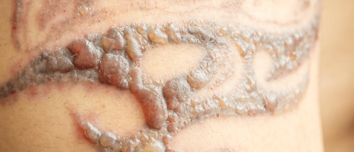
The Role of Chronic Inflammation in Skin Health Post-Tattoo Removal
After laser treatment, the body mounts a typical inflammatory response to the disrupted skin and fragmented ink particles. This acute inflammation is part of the healing process. However, when procedures are repeated frequently or performed improperly, inflammation can become chronic. Chronic inflammation is a well-established precursor to several forms of cancer because it alters cellular repair mechanisms, fosters oxidative stress, and impairs normal immune surveillance.
Some dermatological case reports describe prolonged redness, swelling, or irritation lasting weeks after laser sessions. Histological examinations have occasionally identified elevated levels of cytokines, macrophages, and fibrotic activity in these areas. While this doesn’t imply immediate danger, ongoing inflammatory signaling may theoretically increase the probability of atypical cell changes over time, particularly in genetically predisposed individuals or those with autoimmune conditions.
Therefore, it’s essential to space sessions appropriately, monitor skin response closely, and address any long-term irritation with dermatological consultation to mitigate this risk. Individuals already managing chronic inflammatory conditions should consult with oncologists or immunologists before proceeding with tattoo removal involving large skin areas.
Differences Between Laser Technologies and Their Risk Profiles
Not all lasers are equal when it comes to safety. Tattoo removal primarily employs Q-switched lasers — especially Nd:YAG, ruby, and alexandrite types — that differ in wavelength, penetration depth, and absorption by pigment color. Some systems, like the picosecond laser, offer improved fragmentation efficiency with lower overall energy, which may reduce tissue stress.
Ruby lasers (694 nm) are particularly effective on blue and green inks but can cause higher melanin absorption, posing risks for darker skin tones. Nd:YAG (1064 nm) is safer for deep dermal work and darker skin but less effective for certain pigments. Alexandrite lasers (755 nm) balance efficiency and safety but may still trigger unintended chemical reactions depending on the ink composition.
There is no conclusive evidence that one laser type causes more carcinogenic degradation than another, but variations in energy delivery can impact thermal breakdown of ink. Professionals should match laser systems to pigment type, depth, and patient skin tone — not only for effectiveness, but for minimizing unknown toxicological interactions. As with the treatment of neuroendocrine liver cancer described in the article Stage 4 neuroendocrine cancer spread to liver life expectancy, the choice of technology affects the prognosis and risks
Preventative Strategies to Minimize Long-Term Health Risks
Reducing potential cancer-related risks of tattoo removal starts with informed choices. Patients should research the clinic’s laser systems, demand transparency about ink composition if available, and undergo thorough skin evaluations before beginning the procedure. Using lasers with minimal heat dispersion and shorter pulse durations can reduce unnecessary tissue exposure.
Another consideration involves post-treatment care. Applying anti-inflammatory and antioxidant skin treatments may help neutralize residual reactive compounds. Sun protection is essential, as UV exposure can compound oxidative stress on healing skin. Ensuring a healthy immune system and avoiding known carcinogens like tobacco can further support long-term skin integrity and reduce cumulative risk.
From a systemic viewpoint, the real danger comes not from the laser itself, but from poorly regulated pigments and the cumulative chemical burden of multiple cosmetic interventions. Choosing certified professionals, spacing sessions responsibly, and maintaining general health are the most effective strategies for minimizing long-term complications. In the context of chronic exposure, the article Do parasites cause breast cancer explains how systemic and environmental exposures can interact to influence cancer risk.
Research Gaps and Future Directions in Tattoo Ink Safety
Despite the growth of the tattoo industry and demand for removals, long-term studies evaluating cancer risks remain limited. Most existing data focus on acute reactions or animal models, while large-scale human epidemiological research is scarce. Regulatory agencies such as the FDA and European Chemicals Agency have only recently begun cataloging ink components and their breakdown products.
The need for standardized ink formulation is urgent. Without universal regulations, new inks with unknown chemical behavior continue to enter the market. Furthermore, patient education lags behind the pace of innovation in laser technology and cosmetic practices.
Future directions must include longitudinal studies tracking cancer incidence in individuals who undergo extensive tattooing and laser removal. Multi-disciplinary research involving dermatology, oncology, toxicology, and materials science is essential to develop safer pigments, more targeted laser technologies, and better aftercare protocols. Until then, precaution, transparency, and professional oversight remain the best defense.
FAQ
Can tattoo removal cause cancer directly?
There is no direct scientific proof that laser tattoo removal causes cancer. However, the laser can break down certain ink pigments into potentially carcinogenic byproducts. While these are usually present in small, manageable amounts, repeated exposure may increase theoretical risk in some individuals.
What chemicals are released during laser tattoo removal?
When lasers interact with tattoo ink, they can produce compounds like aromatic amines, benzene derivatives, and reactive oxygen species. These can vary depending on the ink color and composition and are more common in pigments like black or red.
Is laser tattoo removal safe for people with a family history of cancer?
It can be performed safely, but individuals with a genetic predisposition to cancer should consult a medical professional beforehand. The procedure’s effect on skin and immune responses could, in theory, act as a minor additional risk factor.
Does the color of the tattoo matter in cancer risk?
Yes, darker pigments such as black and certain reds contain compounds that are more likely to degrade into potentially harmful byproducts under laser exposure. Lighter colors like yellow may also produce breakdown products of concern.
Are there safer methods than laser for removing tattoos?
Laser removal is currently the most effective and standardized method. Other methods like dermabrasion or surgical excision carry different risks and are less commonly used today.
How can I reduce the risks after tattoo removal?
Post-treatment care is crucial. Avoid sun exposure, apply anti-inflammatory ointments, and follow wound-care instructions. Maintaining a healthy immune system can also help mitigate any long-term risks.
Have there been cancer cases linked to tattoo removal?
To date, there are no confirmed cases where laser tattoo removal was proven to cause cancer in humans. Most evidence is theoretical or based on animal studies.
Are all tattoo inks equally risky?
No. Some inks, especially unregulated ones or those imported without documentation, contain metals and organic compounds with higher toxic potential. The lack of ingredient transparency makes it difficult to assess risk accurately.
Is it safe to remove a tattoo during pregnancy?
It is not recommended. Laser removal involves pain, inflammation, and possible systemic absorption of byproducts, which could affect fetal development. Most clinics advise waiting until after childbirth.
Do older tattoos pose less risk during removal?
Older tattoos often fade and break down naturally, meaning fewer pigment particles may be present. However, the ink may have migrated deeper into the skin or lymphatic system, making removal more complex.
Can laser treatments stimulate dormant cancer cells?
There is no current evidence to support this in the context of tattoo removal. Lasers used in dermatology do not emit ionizing radiation and are unlikely to activate dormant cancer cells.
How is the body affected by broken-down ink particles?
The immune system clears fragmented pigments through macrophages and lymphatic drainage. Some particles may remain in lymph nodes, but there is no solid proof they cause systemic harm.
Does the number of sessions matter in terms of risk?
Yes, more sessions increase cumulative exposure to thermal and chemical changes. While individual risk remains low, frequent treatments on large tattoo areas could compound potential concerns.
What skin reactions should I watch for after removal?
Normal reactions include redness, blistering, and scabbing. If symptoms persist beyond a few weeks or worsen over time, a dermatologist should be consulted to rule out chronic inflammation.
Can people with autoimmune diseases get tattoos removed?
They can, but the risk of abnormal healing or persistent inflammation may be higher. Pre-treatment consultation with a dermatologist and immunologist is strongly advised.