Does a DEXA Scan Show Cancer? Understanding Its Capabilities and Limitations
- What Is a DEXA Scan and How Does It Work?
- What Conditions Are Typically Diagnosed Using a DEXA Scan?
- Can a DEXA Scan Detect Bone Cancer or Metastases?
- Why DEXA Alone Is Not Sufficient for Cancer Diagnosis
- How Bone Metastasis May Be Indirectly Observed on a DEXA Scan
- The Role of DEXA in Cancer Patients Undergoing Treatment
- How DEXA Compares to Other Cancer Imaging Techniques
- When Might a Doctor Suspect Cancer From a DEXA Result?
- Limitations of DEXA Scans in Detecting Malignancy
- Comparative Effectiveness: DEXA vs. Other Imaging Modalities
- When DEXA May Be Incidentally Involved in Cancer Detection
- Clinical Scenarios Where DEXA is Misinterpreted as a Cancer Tool
- Recommendations for Patients at Risk of Cancer
- Role of DEXA in Long-Term Cancer Care
- Clinical Guidelines and Best Practices
- Integrating DEXA Scans into Multimodal Diagnostic Plans
- FAQ

What Is a DEXA Scan and How Does It Work?
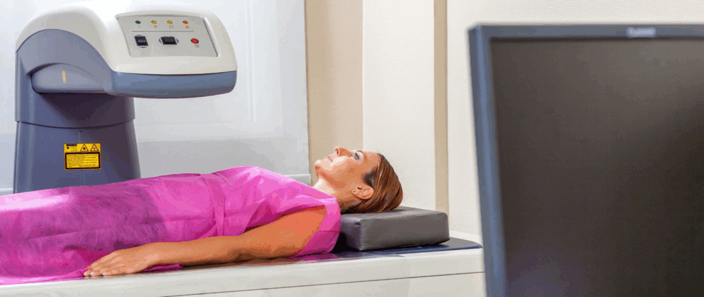
A DEXA scan, also known as dual-energy X-ray absorptiometry, is primarily a diagnostic imaging tool designed to measure bone mineral density. The technique uses two low-dose X-ray beams directed at the bones from different angles, allowing physicians to determine bone loss and assess the risk of osteoporosis. It works on the principle that bone and soft tissue absorb X-rays differently, providing detailed information on bone composition and density. The process is non-invasive, takes approximately 10–30 minutes, and is generally painless. It is considered one of the most accurate methods to evaluate bone strength and fragility, but it is not traditionally classified as a cancer detection tool.
What Conditions Are Typically Diagnosed Using a DEXA Scan?
DEXA scans are almost exclusively used to assess bone-related health issues. Their primary clinical role is in diagnosing osteoporosis, osteopenia, and other metabolic bone disorders. For example, patients who are postmenopausal, on long-term corticosteroids, or have risk factors such as rheumatoid arthritis are routinely evaluated with DEXA. It’s also used for monitoring changes in bone density over time, especially during osteoporosis treatment. Some researchers and clinicians might look at unusual bone density changes as potential red flags for pathological conditions, but in standard practice, the DEXA scan is not used to find or monitor cancer.
Can a DEXA Scan Detect Bone Cancer or Metastases?
DEXA is not intended to detect cancer directly, and it does not differentiate between healthy bone and cancerous bone tissue. However, significant or unusual changes in bone density can occasionally alert physicians to potential issues, including bone metastases. When certain cancers like breast, lung, or prostate cancer spread to the bone, they may cause either osteolytic (bone destroying) or osteoblastic (bone forming) lesions. These abnormalities can sometimes present as atypical findings on a DEXA scan, prompting further diagnostic evaluation. However, more precise imaging such as PET scans, MRIs, or bone scintigraphy are required to identify and characterize cancer involvement accurately.
Why DEXA Alone Is Not Sufficient for Cancer Diagnosis
The sensitivity of a DEXA scan is limited to changes in bone mineral density and structural integrity, not cellular abnormalities. Cancer, especially in early stages, often involves molecular and metabolic changes long before anatomical alterations occur. DEXA lacks the ability to visualize soft tissue tumors or detect malignancies outside of the skeletal system. Furthermore, it cannot distinguish between different causes of bone density changes — for example, whether bone thinning is due to aging, hormonal changes, or metastatic disease. This diagnostic limitation underscores the need for additional imaging and clinical correlation if cancer is suspected. In this context, it becomes evident that modalities like CT, MRI, or PET are superior in evaluating suspicious lesions or cancer progression. Just as a DEXA scan cannot reveal superficial tissue changes like those found in early breast cancer skin mets, it is similarly ineffective for evaluating early soft tissue malignancies.

How Bone Metastasis May Be Indirectly Observed on a DEXA Scan
While DEXA scans are not diagnostic tools for cancer, they can sometimes capture patterns suggestive of underlying metastatic disease, particularly in bones. When cancer cells spread to the skeletal system, they can cause the bone to either degrade or become abnormally dense. These transformations—osteolytic or osteoblastic activity—alter the bone’s mineral density. A sharp decline in BMD (bone mineral density) in a specific region or asymmetrical patterns not typical of osteoporosis may trigger concern. Although DEXA results may raise a clinical suspicion, they cannot confirm a diagnosis or identify the nature of the pathology. Thus, while the scan may hint at bone involvement from a distant malignancy, it is only a preliminary step that must be followed by more targeted oncologic imaging.
The Role of DEXA in Cancer Patients Undergoing Treatment
Patients with cancer, particularly those with breast or prostate malignancies, often undergo therapies—such as chemotherapy, hormone therapy, or radiation—that impact bone health. In these cases, DEXA scans serve an essential purpose in monitoring bone loss and fracture risk rather than detecting the cancer itself. For example, aromatase inhibitors used in breast cancer significantly reduce estrogen levels, accelerating bone thinning. DEXA can track this effect and guide bone-strengthening interventions like bisphosphonates. This monitoring role is crucial for maintaining quality of life in cancer survivors. However, it’s important to distinguish between DEXA’s monitoring function in oncology and its diagnostic limitations regarding cancer detection.
How DEXA Compares to Other Cancer Imaging Techniques
The comparative utility of DEXA versus other imaging modalities used in cancer diagnostics is best understood by examining their intended purposes, strengths, and weaknesses:
| Imaging Technique | Primary Purpose | Can Detect Cancer? | Best For |
| DEXA Scan | Bone density assessment | Indirectly | Monitoring osteoporosis in cancer patients |
| PET Scan | Metabolic activity visualization | Yes | Locating active cancer cells across the body |
| CT Scan | Cross-sectional anatomical imaging | Yes | Tumor size, lymph node involvement |
| MRI | Detailed soft tissue imaging | Yes | Brain, spine, muscles, and soft tissue cancers |
| Bone Scintigraphy | Bone metabolism assessment | Yes | Detecting bone metastases |
This comparison clarifies that while DEXA can be useful in an oncology context, it lacks the resolution and functional specificity needed to serve as a primary cancer detection tool.

When Might a Doctor Suspect Cancer From a DEXA Result?
Although uncommon, there are scenarios in which a doctor might become suspicious of cancer after reviewing a DEXA scan. For example, an unexpected and pronounced decrease in bone density in a young, otherwise healthy individual may prompt questions about an underlying condition, including malignancy. Similarly, isolated regions of abnormally dense bone could raise concern for metastatic disease. These findings would not confirm cancer but would signal the need for further diagnostic evaluation. It’s worth noting that certain cancers cause systemic effects on bone—like multiple myeloma—where bone damage may be widespread, and DEXA might show generalized weakening. Nonetheless, these indications only serve as clues; confirmation always depends on follow-up scans and laboratory work. Interestingly, the same systemic effects that may reduce bone density in DEXA scans can also lead to metabolic imbalances like low phosphate levels, commonly seen in advanced cancers.
Limitations of DEXA Scans in Detecting Malignancy
While DEXA scans are a gold standard for bone density assessment, their use in identifying cancerous lesions comes with inherent limitations. A DEXA scan provides a two-dimensional view of bone mineral content, but it does not offer sufficient resolution or contrast to differentiate between benign and malignant changes in bone. This is particularly true in early cancer stages, where subtle changes might not significantly affect overall bone density. Furthermore, soft tissue abnormalities, which are often the initial indicators of many cancers, are entirely outside the scope of DEXA capabilities. For example, breast or prostate cancers that metastasize to the bone may not present with a detectable reduction in density until the later stages. Additionally, DEXA’s insensitivity to tumor type and aggressiveness means it cannot distinguish between osteoblastic (bone-forming) and osteolytic (bone-destroying) lesions. These factors limit the diagnostic utility of DEXA for cancer detection, reinforcing the need for more advanced imaging techniques when malignancy is suspected.
Comparative Effectiveness: DEXA vs. Other Imaging Modalities
Understanding how DEXA compares to other imaging tools is essential in evaluating its role in cancer diagnostics. The table below summarizes the relative strengths and weaknesses of DEXA compared to other commonly used imaging technologies in oncological practice.
| Imaging Type | Primary Use | Cancer Detection Capability | Soft Tissue Imaging | Radiation Exposure | Cost Efficiency |
| DEXA | Bone density evaluation | Very limited | No | Very low | High |
| CT Scan | Cross-sectional imaging | Moderate to High | Yes | Moderate to High | Moderate |
| MRI | Soft tissue and bone marrow assessment | High | Excellent | None | Low to Moderate |
| PET Scan | Metabolic activity of tissues | Very high | Yes | High | Low |
| X-ray | Basic bone imaging | Low | Limited | Low to Moderate | Very High |
This comparative view clarifies that while DEXA is a safe and cost-effective method for evaluating osteoporosis, it falls short in comprehensive cancer screening compared to more sophisticated imaging techniques like PET or MRI scans.

When DEXA May Be Incidentally Involved in Cancer Detection
Although DEXA is not designed for cancer detection, there are rare circumstances where incidental findings on DEXA scans may prompt further evaluation. For instance, if the scan shows unexpectedly low bone density in a localized area, especially in younger patients or those with no risk factors for osteoporosis, it might raise red flags. Occasionally, subtle asymmetries between bones, unusual curvatures, or abnormal vertebral shapes might lead a radiologist to recommend follow-up imaging. These incidental clues are not diagnostic but could contribute to the early suspicion of metastatic disease, particularly in patients with a known history of cancer. In this context, DEXA serves more as a preliminary trigger for further exploration rather than a definitive diagnostic tool. That said, interpreting such findings demands experience and must be correlated with clinical data, lab work, and more detailed imaging studies. It’s also worth noting that this occasional benefit does not justify relying on DEXA for oncologic purposes in routine practice.
Clinical Scenarios Where DEXA is Misinterpreted as a Cancer Tool
There are clinical settings in which DEXA scan results are mistakenly overvalued in the cancer diagnostic process. This commonly occurs in patients already undergoing cancer treatment who are routinely monitored for bone loss due to therapies such as aromatase inhibitors or corticosteroids. In these cases, a decrease in bone density is a side effect of treatment, not a marker of disease progression. However, patients may misinterpret worsening DEXA results as a sign of advancing cancer. Similarly, in elderly patients, generalized bone thinning detected on a DEXA scan may be mistaken for cancer-related bone involvement when, in fact, it reflects age-related osteoporosis. Misunderstandings may also arise in patients with metastatic cancer history who believe a “normal” DEXA scan means they are free of skeletal disease — a dangerous assumption, given DEXA’s inability to detect focal lesions. Educating both patients and some non-specialist clinicians about these misinterpretations is vital to avoiding false reassurance or unnecessary alarm.
Recommendations for Patients at Risk of Cancer
For patients who have an elevated risk of cancer due to genetic predisposition, lifestyle, or medical history, a DEXA scan should be viewed as a supportive tool rather than a diagnostic endpoint. While it remains an excellent modality for assessing osteoporosis and monitoring bone health in those undergoing cancer treatment, it is not suitable for cancer detection on its own. Patients with risk factors such as a strong family history of cancer, prior cancer diagnoses, unexplained weight loss, fatigue, or persistent localized pain should undergo more specific imaging like MRI, CT, or PET scans. DEXA may be part of a surveillance regimen for skeletal health in these patients, especially when cancer treatments compromise bone integrity, but it must be coupled with clinical vigilance and symptom evaluation. Early and accurate diagnosis depends on comprehensive imaging strategies, not single-modality approaches.
Role of DEXA in Long-Term Cancer Care
Once a patient is diagnosed with cancer, especially those receiving therapies that affect bone metabolism such as chemotherapy, hormone therapy, or radiation, DEXA assumes a vital but secondary role. Its value lies in monitoring the effects of these treatments on bone density and predicting fracture risk. For example, patients with breast or prostate cancer often experience accelerated bone loss due to treatment-induced estrogen or testosterone suppression. In these scenarios, DEXA becomes a long-term monitoring tool, helping oncologists decide when to initiate bisphosphonates or other bone-strengthening agents. Moreover, in survivorship care, DEXA assists in evaluating late complications of treatment and in guiding preventive care strategies aimed at maintaining musculoskeletal health.
Clinical Guidelines and Best Practices
Medical societies such as the American Society of Clinical Oncology (ASCO) and the National Osteoporosis Foundation offer specific guidelines on when and how DEXA should be used in cancer care. These recommendations clarify that DEXA is not indicated for primary cancer detection. Instead, it is recommended for patients starting endocrine therapies, postmenopausal women with cancer, and individuals with prolonged corticosteroid use. Clinical best practices also emphasize combining DEXA findings with laboratory markers of bone turnover, calcium and vitamin D levels, and patient-reported symptoms to create a comprehensive picture. Additionally, follow-up scans should be scheduled based on treatment duration and baseline bone health rather than arbitrary intervals. Clear communication of the DEXA scan’s purpose is also part of best practices to avoid patient confusion regarding its diagnostic limits.
Integrating DEXA Scans into Multimodal Diagnostic Plans
DEXA scans should never be interpreted in isolation when cancer is suspected or being monitored. Their optimal use occurs when integrated into a multimodal diagnostic framework that includes clinical evaluation, blood tests, and other imaging modalities. In a well-structured cancer diagnostic plan, DEXA contributes essential data about skeletal health and fracture risk but must be interpreted in the context of the broader clinical picture. For instance, in patients with back pain and a known history of malignancy, combining DEXA with spinal MRI provides both bone density and structural insights. Similarly, patients undergoing PET/CT scans for tumor mapping may use DEXA concurrently to assess treatment impact on bone. This integration ensures that while DEXA doesn’t detect tumors, it supports holistic cancer management. As in the article on whether low phosphate levels can cause cancer, it is important to consider the metabolic changes that accompany cancer processes.
FAQ
What is a DEXA scan used for?
A DEXA (Dual-Energy X-ray Absorptiometry) scan is primarily used to measure bone mineral density. It is a widely accepted method for diagnosing osteoporosis and assessing fracture risk, especially in postmenopausal women and older adults. In cancer patients, it also helps monitor the effects of treatments that may weaken bones.
Can a DEXA scan detect cancer?
No, a DEXA scan cannot directly detect cancer. Its function is to evaluate bone density, not to identify tumors, abnormal tissue growth, or cancerous lesions. For cancer diagnosis, imaging techniques like CT, MRI, or PET scans are required.
Why might a cancer patient need a DEXA scan?
Cancer patients may need a DEXA scan to monitor their bone health, especially if they are undergoing treatments such as chemotherapy, hormone therapy, or corticosteroids. These treatments can reduce bone density, and a DEXA scan helps assess the risk of fractures.
What are the limitations of a DEXA scan in oncology?
DEXA scans cannot differentiate between healthy and malignant bone tissue, identify soft tissue tumors, or detect metastatic spread. They are limited to assessing bone mineral density and cannot substitute for diagnostic cancer imaging.
Is a DEXA scan safe for cancer patients?
Yes, DEXA scans are considered very safe. They use very low doses of radiation, significantly lower than standard X-rays or CT scans. This makes them appropriate even for patients requiring repeated assessments over time.
Can a DEXA scan show bone metastases?
DEXA scans cannot reliably show bone metastases. While metastatic cancer may lead to bone loss that shows up as reduced density, DEXA cannot confirm the presence or type of underlying malignancy. More advanced imaging is necessary for detection.
How is DEXA used during cancer treatment?
During cancer treatment, DEXA is used to monitor the impact of therapies on bone mass. Oncologists use it to detect early signs of osteoporosis and make decisions about bone-strengthening medications or lifestyle modifications.
Do all cancer patients need a DEXA scan?
Not all cancer patients require a DEXA scan. It is generally recommended for those receiving treatments known to affect bone health, such as hormone-blocking therapies or long-term corticosteroids. It may also be advised for older patients or those at risk of osteoporosis.
How often should cancer patients have a DEXA scan?
The frequency of DEXA scans depends on the patient’s baseline bone density, the type of cancer treatment they are receiving, and their overall fracture risk. Typically, repeat scans are done every 1–2 years, but high-risk cases may require more frequent monitoring.
What does a low DEXA score mean for a cancer patient?
A low DEXA score in a cancer patient indicates reduced bone density and an increased risk of fractures. It often necessitates a treatment plan that may include calcium and vitamin D supplementation, lifestyle changes, and possibly medications like bisphosphonates.
Can DEXA scans help predict complications during cancer treatment?
Yes, by identifying low bone density early, DEXA scans can help anticipate complications such as fractures, especially in the spine or hips. This allows for timely interventions that can improve patient outcomes and reduce treatment-related morbidity.
Does insurance cover DEXA scans for cancer patients?
Insurance often covers DEXA scans for cancer patients if there is a documented risk of bone loss or osteoporosis due to treatment. Coverage may vary depending on individual plans, the patient’s age, and clinical indications provided by the physician.
Is DEXA used in cancer screening programs?
No, DEXA is not part of standard cancer screening protocols. Its role is focused on bone health assessment and is not appropriate for detecting or screening for cancer. Mammograms, colonoscopies, and low-dose CT scans are typical tools for cancer screening.
What symptoms would warrant imaging beyond DEXA?
Symptoms such as persistent bone pain, unexplained fractures, weight loss, or neurological signs should prompt further imaging like MRI or CT, as these may indicate cancer involvement that DEXA cannot detect.
How does DEXA compare to PET scans for cancer detection?
DEXA and PET scans serve entirely different purposes. DEXA evaluates bone density, while PET scans detect metabolic activity and can identify cancerous tissue. PET scans are superior for locating and staging cancer, whereas DEXA supports bone health assessment during or after treatment.


