Breast Cancer Spreading to the Skin: Symptoms and Treatment Options
- What Does It Mean When Breast Cancer Spreads to the Skin?
- How Breast Cancer Cells Reach the Skin: Mechanisms and Pathways
- Visual Appearance and Symptom Variability in Skin Metastases
- Tools and Techniques for Confirming Diagnosis
- Histological Characteristics of Breast Cancer Skin Metastases
- Breast Cancer Subtypes and Their Relationship to Skin Spread
- Prognostic Implications of Skin Metastases
- Treatment Strategies: Combining Local and Systemic Approaches
- Side Effects and Challenges of Treating Skin Metastases
- Emotional and Psychological Impact on Patients
- Skin Metastases in Rare Sites: Scalp, Face, and Extremities
- Follow-up Care and Monitoring of Skin Lesions
- Long-Term Survival and Quality of Life with Skin Metastases
- Cutaneous vs Other Common Breast Cancer Metastasis Sites
- Special Considerations in Recurrent or Treatment-Resistant Lesions
- Preventive Measures and Patient Education
- Frequently Asked Questions
What Does It Mean When Breast Cancer Spreads to the Skin?
When breast cancer spreads to the skin, it usually signifies an advanced or metastatic stage of the disease, indicating that cancer cells have moved beyond the primary breast tissue and established colonies in the dermal or subdermal layers. This process is known medically as “cutaneous metastasis.” In breast cancer, the skin is among the more frequent sites of secondary tumor development, especially in patients with a high systemic tumor burden or aggressive subtypes like inflammatory breast cancer. It does not necessarily mean the skin is the only organ affected—rather, it often reflects more widespread internal dissemination. These skin manifestations might appear months or even years after the initial diagnosis or treatment, underscoring the importance of long-term monitoring. The visual changes can range from subtle discolorations to extensive ulcerations, all of which can significantly affect the patient’s quality of life and prognosis. Cutaneous involvement generally signals the need for systemic reevaluation and therapeutic adjustment.
How Breast Cancer Cells Reach the Skin: Mechanisms and Pathways
Breast cancer cells can reach the skin through two main biological routes: lymphatic spread and hematogenous dissemination. In the lymphatic route, malignant cells travel through lymph vessels, which are abundant around the breast and chest wall, eventually infiltrating the superficial dermis. This mechanism is particularly common in locally advanced cancers and explains why many cutaneous lesions develop near surgical scars or radiation fields. Hematogenous spread involves cancer cells entering the bloodstream and being transported to distant skin sites, including the scalp, face, or extremities—areas not typically associated with the primary tumor. Once lodged in the skin’s microvasculature, these cells begin to divide and form palpable nodules or infiltrative plaques. Inflammatory breast cancer, a highly aggressive subtype, is known for diffuse skin involvement that mimics cellulitis or dermatitis, creating diagnostic challenges. Understanding these pathways is crucial not only for diagnosis but also for anticipating possible recurrence patterns and tailoring surveillance.
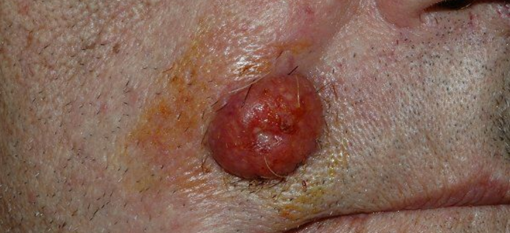
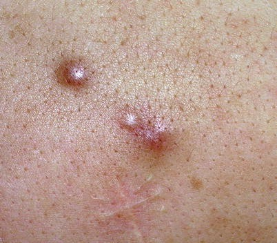
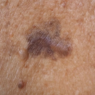
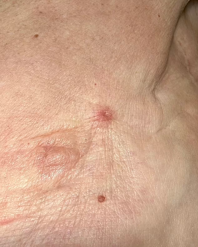
Visual Appearance and Symptom Variability in Skin Metastases
The cutaneous manifestations of metastatic breast cancer are notoriously variable, often depending on tumor biology, patient immune response, and previous treatments like radiation or surgery. Skin lesions may present as firm, skin-colored or reddish nodules that grow slowly or rapidly. In some patients, these nodules coalesce into plaques that cover large areas of the chest or abdomen, while in others, they may ulcerate or crust, creating an open wound that is prone to infection. Less commonly, patients may develop telangiectatic patches, eczema-like rashes, or even blister-like lesions, mimicking benign conditions such as herpes zoster or fungal infections. Pain is not always present; some lesions may be entirely asymptomatic and only discovered during routine examination. However, others may produce discomfort, itching, burning, or a tightening sensation, especially when they cause significant dermal thickening or edema. Because these skin changes can be subtle and non-specific, any persistent lesion in a patient with a breast cancer history must be evaluated thoroughly. Please note that some hormonal therapies, including Sermorelin, may complicate skin testing due to altered tissue response.
Tools and Techniques for Confirming Diagnosis
Accurate diagnosis of skin metastases requires a combination of visual inspection, patient history, and confirmatory pathological testing. While clinical appearance offers initial clues, definitive diagnosis rests on biopsy, preferably an incisional or punch biopsy, which allows for examination of full dermal architecture. The pathologist will evaluate cellular morphology, presence of ductal structures, mitotic activity, and use immunohistochemical markers such as estrogen receptor (ER), progesterone receptor (PR), HER2, and cytokeratin 7 to confirm breast origin. Imaging studies, although less specific for skin lesions, help determine the full extent of metastasis and detect deeper tissue involvement. In some cases, a PET-CT scan is ordered to assess for concurrent internal organ involvement. Dermoscopy can be helpful in identifying vascular patterns or irregular pigmentation, although it’s not typically diagnostic on its own. If patients are undergoing treatment for other conditions, such as osteoporosis or hormone imbalance, it is crucial to consider the interaction with systemic disease—a good example being how a DEXA scan might reveal signs of metastatic bone lesions, even if initial suspicion is unrelated.
Histological Characteristics of Breast Cancer Skin Metastases
Histological analysis of skin metastases reveals patterns that closely resemble the primary tumor’s architecture. In breast cancer, the metastatic skin lesions often consist of infiltrating ductal carcinoma cells arranged in nests, cords, or sheets within the dermis. These cells may appear pleomorphic, with prominent nucleoli and high mitotic activity, especially in high-grade tumors. The presence of glandular formations or mucin secretion may further support the diagnosis. One notable feature is the absence of epidermal involvement in early lesions—metastatic cells typically spare the outermost layer of the skin and instead localize deeper, within the dermal and subcutaneous layers. Immunohistochemical staining plays a crucial role in confirming the diagnosis: most skin metastases from breast cancer are positive for cytokeratin 7, ER/PR, and mammaglobin. HER2 status should also be evaluated, as it influences treatment decisions. Importantly, histology not only confirms the origin but also helps distinguish between cutaneous metastases and primary skin cancers such as basal or squamous cell carcinoma.
Breast Cancer Subtypes and Their Relationship to Skin Spread
Different subtypes of breast cancer carry varying risks and patterns of skin involvement. Inflammatory breast cancer is the most well-known for early and aggressive cutaneous spread, often presenting as redness, swelling, and warmth over large areas of the chest wall—a presentation that mimics infection but is actually caused by cancer cells blocking lymphatic vessels. Triple-negative breast cancer (TNBC), although less likely to cause diffuse skin involvement, can lead to nodular skin metastases due to its aggressive and fast-growing nature. HER2-positive tumors are also associated with skin spread, particularly in resistant cases where targeted therapies have failed. Hormone receptor-positive subtypes may cause skin metastases years after the original diagnosis, highlighting the need for long-term vigilance even in “low-risk” patients. Each subtype behaves differently in terms of growth speed, treatment response, and recurrence pattern, and understanding these nuances allows clinicians to predict which patients are most likely to develop cutaneous involvement.
Prognostic Implications of Skin Metastases
The presence of skin metastases in breast cancer is generally considered a sign of systemic disease and indicates stage IV, or metastatic, breast cancer. However, the specific impact on prognosis depends on several factors, including the subtype of breast cancer, the number and size of skin lesions, the presence of other metastatic sites, and overall treatment response. Patients with isolated skin metastases and good performance status may live for years with appropriate systemic therapy and local control measures. On the other hand, widespread cutaneous involvement often reflects a high tumor burden and poor treatment response. Additionally, recurrent skin metastases after complete remission suggest tumor resistance, a common challenge in hormone-refractory and HER2-positive patients. Prognosis must be individualized, but in general, cutaneous spread significantly alters the course of the disease. Just as a DEXA scan can occasionally reveal unsuspected signs of metastatic activity, seemingly localized skin lesions can hint at broader disease progression that requires systemic reassessment.
Treatment Strategies: Combining Local and Systemic Approaches
Treating skin metastases from breast cancer requires a multimodal strategy that addresses both the visible skin lesions and the underlying systemic disease. Local treatments such as surgical excision, cryotherapy, or topical agents (e.g., imiquimod or 5-fluorouracil) may be used for isolated or symptomatic lesions but are rarely curative on their own. Radiation therapy is particularly useful in controlling bleeding, pain, or rapid lesion growth and can be delivered as part of palliative care or alongside systemic treatment. Systemic therapy remains the cornerstone of management. This includes endocrine therapy for hormone receptor-positive cancers, HER2-targeted agents like trastuzumab or pertuzumab for HER2-positive cases, and chemotherapy for triple-negative or progressive disease. Immunotherapy is an emerging option, especially in PD-L1-positive TNBC. The chosen regimen must reflect the tumor’s biology, treatment history, and the patient’s overall health. Skin response is often a visible marker of systemic treatment effectiveness, and reduction in skin lesions can indicate broader disease control.
Side Effects and Challenges of Treating Skin Metastases
Treatments for breast cancer skin metastases—though essential—often come with substantial side effects and management challenges. Radiation therapy, while effective for localized control, can lead to skin irritation, blistering, and delayed healing, particularly in areas previously irradiated during initial breast cancer treatment. Chemotherapy and targeted therapies can cause generalized skin dryness, photosensitivity, or dermatologic toxicity, which may worsen the patient’s condition. When immunotherapy is part of the regimen, immune-related skin reactions such as rash or pruritus may develop and sometimes require corticosteroids or treatment suspension. Another key issue is superinfection—open or ulcerated skin metastases create a portal for bacterial entry, potentially leading to cellulitis or even sepsis. Management includes balancing oncologic efficacy with skin protection, using barrier creams, systemic antibiotics, and sometimes wound care consultations. Quality of life may be severely affected by cosmetic disfigurement or persistent discomfort. Physicians must work closely with dermatologists, palliative care teams, and wound care specialists to ensure holistic care.
Emotional and Psychological Impact on Patients
The psychological toll of visible metastases cannot be overstated. Patients who experience skin involvement often report feelings of anxiety, social withdrawal, and fear of stigma due to the visibility of their disease. For many, skin lesions are a constant visual reminder of their cancer’s progression, leading to distress and disrupted body image. This is particularly true for women who have undergone mastectomy and now face recurrent disease in the same region, causing emotional setbacks. Open, draining, or ulcerating lesions can impact intimacy and reduce self-confidence, leading to depression or avoidance behavior. Supportive psychotherapy, patient education, and peer support groups are essential tools in managing this aspect of care. Health care providers should proactively address these issues and encourage patients to express concerns. Even when lesions are medically manageable, the visible nature of skin metastases requires sensitive handling and empathetic communication from the care team.
Skin Metastases in Rare Sites: Scalp, Face, and Extremities
Although the chest wall is the most common site for cutaneous metastases in breast cancer, less typical locations can also be affected, including the scalp, face, and extremities. Metastases in these areas may be mistaken for dermatologic conditions such as folliculitis, eczema, or benign cysts, delaying diagnosis. The scalp is a particularly challenging area because hair can obscure lesions, and biopsies in this region require careful technique. Facial metastases can be emotionally and socially distressing, as they are difficult to conceal and often associated with a worse prognosis due to concurrent visceral spread. Lesions on the limbs may present as nodules or plaques and are often overlooked, especially in patients with widespread disease. Their detection, however, can be vital for assessing treatment response or disease progression. The rarity of these locations underscores the need for vigilance in full-body skin exams, especially when patients report new symptoms. Just as mucoceles are sometimes mistaken for harmless growths, skin metastases on the face or head are often diagnosed late due to their unusual location.
Follow-up Care and Monitoring of Skin Lesions
After the initial diagnosis and treatment of breast cancer skin metastases, follow-up care is essential to monitor therapeutic response and detect early signs of progression or recurrence. Dermatologic evaluation should be part of routine oncology follow-up, particularly in patients with previous cutaneous involvement. Physicians should document lesion size, number, appearance, and symptom evolution at each visit. Serial photography can assist in visual comparisons over time. If new skin changes arise or existing lesions worsen, biopsy may be repeated to rule out new primary tumors or resistance-driven histological changes. Systemic imaging, including PET or CT, may be needed to evaluate for deeper or internal progression. Additionally, skin complications such as infections, necrosis, or lymphedema may require interdisciplinary management. Continuous follow-up ensures that treatment regimens remain aligned with the patient’s evolving condition, and any dermatologic emergencies are addressed promptly.
Long-Term Survival and Quality of Life with Skin Metastases
Although skin metastases from breast cancer generally signal advanced disease, they do not always equate to imminent death. With modern systemic therapies, many patients live for months or even years, maintaining a reasonable quality of life. Treatment advances have allowed for better symptom control, reduced lesion burden, and longer progression-free intervals. Hormone therapies, targeted biologics, and immunotherapies have shown particular promise in managing slow-progressing or receptor-positive tumors. That said, the visibility of skin metastases—especially on the chest wall or face—can impose significant psychological and physical burdens. Long-term survival depends on cancer subtype, patient age, response to treatment, and presence of internal metastases. While curative outcomes are rare, palliative treatment can achieve meaningful results, giving patients time, comfort, and dignity.
Cutaneous vs Other Common Breast Cancer Metastasis Sites
| Feature | Skin Metastases | Bone Metastases | Lung Metastases | Liver Metastases |
| Frequency in Advanced Cases | Moderate (~23–25%) | High (~60–70%) | Moderate (~30–40%) | Moderate (~30–40%) |
| Common Symptoms | Nodules, rash, ulceration | Pain, fractures, spinal issues | Cough, shortness of breath | Jaundice, abdominal swelling |
| Diagnostic Method | Skin biopsy | Bone scan, DEXA, MRI | Chest CT, PET scan | Liver function tests, MRI |
| Prognostic Indicator | Suggests systemic progression | Early marker in ER+ cancers | Often follows bone spread | Often seen with aggressive subtypes |
| Management | Local + systemic therapy | Endocrine + bisphosphonates | Chemotherapy ± targeted agents | Chemotherapy ± liver-directed therapy |
This table summarizes key differences in behavior, detection, and treatment strategies for breast cancer metastases at different sites, including the skin. Each location has unique implications for prognosis and care planning.
Special Considerations in Recurrent or Treatment-Resistant Lesions
In some patients, skin metastases persist or recur despite standard treatment protocols, highlighting the need for personalized care plans. Resistance may develop through mutations in estrogen receptor pathways, HER2 amplification, or tumor microenvironment adaptation. Molecular profiling of biopsy specimens can help guide second-line or third-line therapy options. For instance, PI3K inhibitors may be effective in certain resistant hormone receptor-positive tumors, while PARP inhibitors are valuable in BRCA-mutated cases. Refractory lesions may also respond to palliative radiation or electrochemotherapy, a newer approach combining chemotherapy with electric pulses to improve cell uptake. Clinical trials offer another avenue, especially for patients whose tumors express rare or novel markers. Importantly, multidisciplinary coordination between oncology, dermatology, and palliative care is vital to ensure all possible options are considered and quality of life remains a central focus.
Preventive Measures and Patient Education
Although cutaneous metastases cannot always be prevented, early recognition and patient education can dramatically improve outcomes. Breast cancer patients should be taught to monitor their skin regularly, particularly near surgical scars, the chest wall, or radiation sites. Any new or persistent skin lesion, especially those that are firm, discolored, or bleeding, must be brought to medical attention promptly. Routine oncologic follow-ups should include full-body skin assessments. Healthcare providers must also ensure that patients understand the signs of recurrence and are equipped to report changes between appointments. Empowering patients through knowledge helps avoid delays in diagnosis and intervention, which in turn may slow progression and alleviate unnecessary suffering. Good communication, visual documentation of lesions, and access to rapid dermatologic referral systems play a vital role in proactive management.
Frequently Asked Questions
What does it mean when breast cancer spreads to the skin?
When breast cancer spreads to the skin, it indicates that malignant cells have traveled beyond the original tumor site and formed secondary lesions in the dermal layers. This condition, known as cutaneous metastasis, signifies systemic disease progression and generally classifies the cancer as stage IV.
How do skin metastases from breast cancer look?
These lesions often appear as firm, painless nodules, reddish or purplish plaques, ulcerated wounds, or areas of thickened, inflamed skin. They may resemble infections, allergic reactions, or benign growths, which can delay accurate diagnosis if not carefully evaluated.
Is skin metastasis a sign of terminal breast cancer?
While skin metastasis signals advanced cancer, it is not necessarily terminal. Many patients respond well to systemic therapies, and with ongoing treatment, they can live for extended periods. The prognosis depends on tumor biology, treatment response, and presence of other metastases.
Can skin metastases be the first sign of breast cancer?
Although uncommon, in some cases, cutaneous metastases may appear before a breast cancer diagnosis, especially in aggressive subtypes. These patients typically present with rapidly growing skin lesions that prompt further oncologic investigation.
Are skin metastases painful?
Not always. Many lesions are painless at first but can become painful due to ulceration, infection, or rapid expansion. Some patients may experience itching, burning, or sensitivity in the affected area, particularly if lesions are exposed to friction.
How are breast cancer skin metastases diagnosed?
Diagnosis involves clinical examination, followed by a biopsy of the skin lesion. Pathological analysis, including immunohistochemical staining, confirms whether the cells originated from breast tissue. Imaging may be used to assess systemic spread.
Can a skin lesion be mistaken for metastasis?
Yes, several benign skin conditions like cysts, infections, or eczema can mimic cutaneous metastases. This is why biopsy confirmation is crucial. Even conditions like mucocele cancer symptoms can sometimes be confused with metastatic behavior when they appear in unusual facial areas.
How are skin metastases treated?
Treatment typically combines local methods such as radiation or minor excision with systemic therapies like hormone treatments, chemotherapy, targeted therapy, or immunotherapy. The goal is to manage symptoms, control growth, and improve overall disease control.
Do skin metastases always mean the cancer is widespread?
Usually, yes. Skin involvement often correlates with metastasis to other organs such as bones, lungs, or liver. However, in rare cases, skin may be the only site of spread for a prolonged period, especially if caught early and treated effectively.
Is radiation effective for skin metastases?
Radiation therapy is highly effective in reducing symptoms such as bleeding, pain, or rapid tumor growth. It may be used alone or in combination with systemic treatment, particularly in patients with localized but symptomatic skin lesions.
Can you live with skin metastases for years?
Yes, some patients—particularly those with slow-growing, hormone receptor-positive tumors—can live for several years with good systemic control. Modern therapies have extended survival significantly, even for those with visible skin lesions.
How often should patients with skin metastases be monitored?
Follow-up schedules depend on disease status and treatment response, but typically involve clinical skin exams every 1–3 months and imaging every 3–6 months. Any changes in skin appearance should prompt immediate reassessment.
What is the risk of recurrence after treatment?
Recurrence is common, particularly in aggressive subtypes or patients with previous widespread disease. New skin lesions often indicate systemic progression and may require changes in therapy. Continuous monitoring is essential.
Do hormonal therapies help with skin metastases?
Yes, for hormone receptor-positive cancers, endocrine therapy is often the first line of treatment and can effectively reduce or stabilize skin metastases. However, resistance may develop over time, necessitating a shift to other modalities.
Can skin metastases affect mental health?
Absolutely. The visibility and chronic nature of skin metastases can lead to anxiety, depression, and reduced self-esteem. Supportive care, counseling, and peer networks are vital in addressing the emotional impact of this condition.