Breast Cancer: Understanding Skin Metastases
- What Are Skin Metastases in Breast Cancer?
- Incidence and Epidemiology
- Clinical Presentation and Diagnosis
- Pathophysiology of Skin Metastases
- Histological Subtypes and Their Implications
- Treatment Strategies
- Importance of Multidisciplinary Care
- Comparative Insights: Skin Metastases in Other Cancers
- Patterns of Spread and Anatomical Sites of Involvement
- Prognostic Implications and Survival Outcomes
- Diagnostic Challenges and Differential Diagnoses
- Integration of Palliative and Supportive Care
- Resistance to Treatment and Disease Progression
- Psychosocial Burden and Body Image Concerns
- Role of Multidisciplinary Teams in Managing Cutaneous Metastases
- Rare and Atypical Presentations of Skin Metastases
- The Importance of Patient Education and Self-Monitoring
- Common Mistakes and Delayed Recognition
- Family Support, Prognostic Conversations, and Advanced Care Planning
- Compassionate Oncology in Visible Disease
- FAQ
What Are Skin Metastases in Breast Cancer?
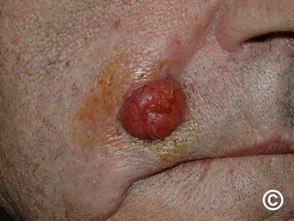
Skin metastases, or cutaneous metastases, occur when breast cancer cells spread from the primary tumor to the skin. This manifestation indicates an advanced stage of cancer, often associated with systemic dissemination. The skin involvement can present in various forms, including nodules, plaques, or inflammatory lesions, and is a sign of disease progression.
Incidence and Epidemiology
Breast cancer is the most common malignancy leading to skin metastases in women. Studies indicate that cutaneous metastases occur in approximately 23.9% to 30% of breast cancer cases . These metastases often develop months or even years after the initial diagnosis and treatment of the primary tumor, highlighting the need for ongoing vigilance in long-term cancer care.
Clinical Presentation and Diagnosis
The clinical presentation of skin metastases varies, often mimicking benign skin conditions, which can delay diagnosis. Common manifestations include:
- Firm, painless nodules
- Erythematous patches resembling cellulitis
- Ulcerative lesions
- Inflammatory plaques
Diagnosis is confirmed through skin biopsy and histopathological examination, which reveal malignant cells consistent with breast carcinoma. Imaging studies may be employed to assess the extent of systemic disease.
Pathophysiology of Skin Metastases
The development of skin metastases involves the dissemination of cancer cells via lymphatic or hematogenous routes. These cells infiltrate the dermis and subcutaneous tissues, leading to the formation of metastatic lesions. The skin’s rich vascular and lymphatic networks provide pathways for tumor cell migration and colonization.
Histological Subtypes and Their Implications
Breast cancer encompasses various histological subtypes, each with distinct behaviors and propensities for skin metastasis. Understanding these subtypes aids in prognostication and treatment planning.
| Histological Subtype | Skin Metastasis Tendency | Prognostic Implications |
| Invasive Ductal Carcinoma | High | Variable |
| Invasive Lobular Carcinoma | Moderate | Often late recurrence |
| Inflammatory Breast Cancer | High | Aggressive behavior |
| Triple-Negative Breast Cancer | High | Poor prognosis |
| HER2-Positive Breast Cancer | Moderate | Responsive to targeted therapy |
Treatment Strategies
Management of skin metastases in breast cancer is multifaceted, focusing on systemic control and local symptom relief. Treatment options include:
- Systemic Therapy: Chemotherapy, hormonal therapy, and targeted agents address the underlying disease.
- Radiation Therapy: Effective for localized skin lesions, providing symptom palliation.
- Surgical Intervention: Reserved for isolated lesions causing significant discomfort or functional impairment.
- Topical Treatments: Agents like imiquimod have shown efficacy in certain cases .
Emerging therapies, such as electrochemotherapy and photodynamic therapy, are under investigation for their potential benefits in managing cutaneous metastases .
Importance of Multidisciplinary Care
Effective management of breast cancer skin metastases requires a multidisciplinary approach, involving oncologists, dermatologists, surgeons, radiation therapists, and palliative care specialists. Collaborative care ensures that treatment plans are tailored to the patient’s needs, optimizing outcomes and quality of life.
Comparative Insights: Skin Metastases in Other Cancers
Skin metastases are not exclusive to breast cancer. For instance, nasal cancer in dogs can also present with cutaneous lesions, emphasizing the importance of recognizing skin involvement across different species and cancer types. Similarly, lower eyelid cancer may manifest with skin changes that require prompt evaluation and management. Understanding these parallels enhances our approach to diagnosing and treating skin metastases across various contexts.
Patterns of Spread and Anatomical Sites of Involvement
Skin metastases from breast cancer often follow a predictable distribution pattern. The most common locations are the chest wall, especially over the site of previous mastectomy or lumpectomy. However, lesions may also appear on the scalp, abdomen, upper arms, or even distant sites like the thighs. The proximity to lymphatic pathways explains this, as tumor cells often travel via superficial lymphatics to dermal tissues.
Sometimes, skin metastases appear as the first sign of systemic recurrence, especially in aggressive subtypes like inflammatory breast cancer or triple-negative breast cancer. These cases underscore the importance of regular skin examinations — even years after initial remission — particularly for patients who received chest wall radiation or extensive lymph node dissection.
Understanding these patterns helps clinicians differentiate recurrence from dermatologic conditions, such as eczema or lymphedema-related skin changes, which can visually mimic cutaneous metastases.
Prognostic Implications and Survival Outcomes
The development of cutaneous metastases is generally associated with advanced disease and a poorer prognosis. However, outcomes can vary significantly depending on the cancer subtype, overall burden of metastatic disease, and the patient’s response to systemic treatment.
In patients with limited skin involvement and otherwise controlled metastases, especially those on effective HER2-targeted or hormonal therapies, progression may be slow, and survival can extend well beyond one year. Conversely, widespread dermal infiltration often signals systemic failure and a median survival closer to 6–9 months.
Interestingly, cutaneous metastases may appear alongside peritoneal spread in aggressive breast cancer subtypes — a metastatic pattern also seen in Stage 4 appendix cancer, where mucinous deposits on the peritoneum often coincide with other organ involvement.
While cutaneous lesions themselves rarely cause death, they signal biologic aggressiveness and necessitate coordinated, multidisciplinary response.
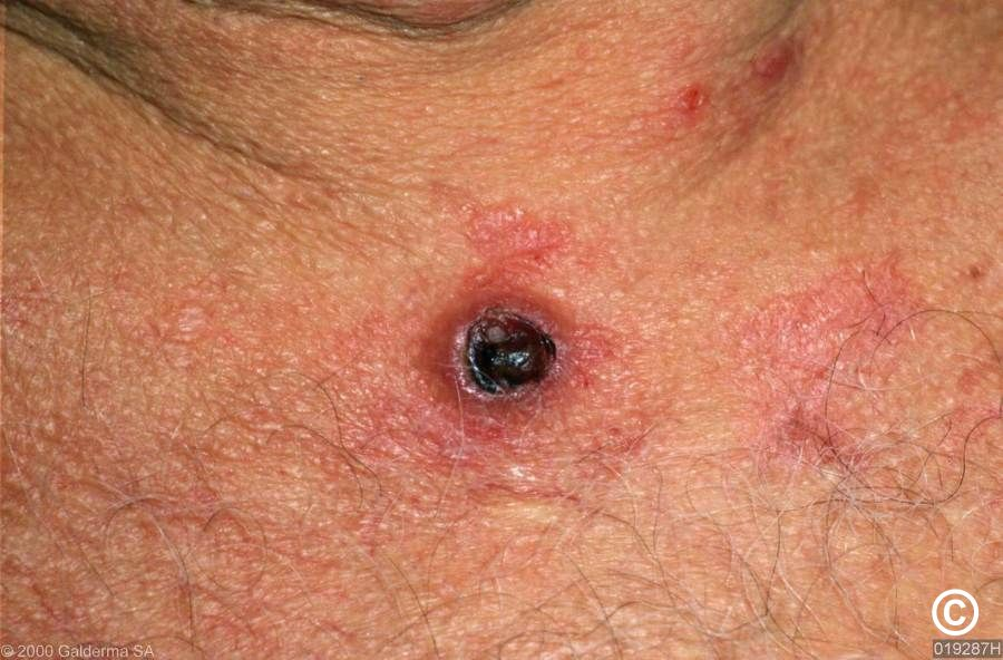
Diagnostic Challenges and Differential Diagnoses
Skin metastases can be deceptively subtle and are frequently misdiagnosed in early stages. Because they may resemble cellulitis, dermatitis, or scar hypertrophy, clinicians must maintain a high index of suspicion in patients with a history of breast cancer who present with new or evolving skin changes.
A punch biopsy is the gold standard for confirming diagnosis. Histology will reveal malignant epithelial cells infiltrating the dermis or lymphovascular spaces, often forming nests or sheets. Immunohistochemistry may be used to confirm hormone receptor or HER2 status, which is essential for guiding systemic therapy.
In rare cases, lesions may ulcerate, bleed, or become infected, leading to diagnostic confusion. This reinforces the importance of dermatologic consultation in any ambiguous presentation.
Integration of Palliative and Supportive Care
Palliative care plays a crucial role in managing patients with breast cancer skin metastases, especially those with widespread lesions, persistent discomfort, or limited systemic options. Early integration of palliative teams helps manage not only physical symptoms like pain, itching, and exudate but also emotional and existential distress.
Symptom-directed interventions may include topical lidocaine, corticosteroids, or specialized wound dressings. For infected or weeping lesions, gentle hygiene, low-adherent dressings, and antibiotics may be necessary. Some patients may also benefit from radiation therapy aimed solely at symptomatic cutaneous nodules.
Palliative teams also help guide conversations about care goals, resuscitation preferences, and the patient’s vision for quality of life — themes that are central in other terminal diagnoses such as Stage 4 appendix cancer and advanced skin-based malignancies like lower eyelid cancer.
Resistance to Treatment and Disease Progression
One of the most challenging aspects of managing cutaneous metastases is therapeutic resistance. Skin metastases may appear even in patients on ongoing systemic therapy, signaling adaptation or mutation within tumor cells. For instance, patients receiving hormonal therapy may develop hormone receptor-negative skin lesions, reflecting clonal evolution.
Progression can also occur during chemotherapy or after initial response to HER2-targeted agents. Biopsy and receptor reassessment are crucial in these cases, as the biology of the metastatic lesion may differ from the primary tumor. Switches in treatment strategy — such as moving to antibody-drug conjugates or immunotherapy (in triple-negative breast cancer) — are considered based on current molecular profiles.
Ultimately, progression of skin metastases often correlates with systemic decline, making real-time monitoring and regular oncology follow-ups essential to adjust care in a timely manner.
Psychosocial Burden and Body Image Concerns
Beyond the clinical burden, cutaneous metastases exert a profound emotional and psychological toll. Because these lesions are visible and often located on the chest, arms, or neck, they can trigger feelings of shame, vulnerability, and altered body identity. Patients frequently report discomfort when dressing, bathing, or appearing in public — especially when lesions are ulcerated or exudative.
For some, the appearance of skin metastases feels like a visible reminder of disease recurrence, reinforcing existential fears and disrupting the fragile balance they’ve built during remission. This emotional impact requires targeted support from oncology psychologists, social workers, and peer counselors.
Group support environments — either in-person or virtual — offer validation, coping strategies, and a safe space for expressing appearance-related grief. While the oncology team focuses on medical care, mental health professionals ensure the patient feels seen beyond the diagnosis.
Role of Multidisciplinary Teams in Managing Cutaneous Metastases
Effective care for patients with skin metastases hinges on seamless coordination between disciplines. Oncologists direct systemic treatment and imaging surveillance, while dermatologists help diagnose, manage symptoms, and track skin-specific progression. Radiation oncologists offer targeted palliation, and surgical teams may assist with debridement or reconstruction in selected cases.
Wound care specialists are indispensable when lesions become necrotic, infected, or ulcerated. They guide the choice of dressings, ensure hygiene, and train families in home-based skin care routines.
This collaboration mirrors the approach used in rare but complex cases like nasal cancer in dogs, where veterinary oncologists, radiologists, and palliative specialists must coordinate to preserve both survival and comfort.
When healthcare providers function as a unified team, patients benefit from faster intervention, clearer communication, and a more personalized therapeutic experience.
Rare and Atypical Presentations of Skin Metastases
While most cutaneous breast cancer metastases appear as nodules or plaques, there are less common patterns that complicate diagnosis. These include:
- Telangiectatic carcinomatosis: Resembling vascular malformations or spider veins
- Carcinoma en cuirasse: Diffuse, hardened skin mimicking scleroderma
- Zosteriform metastases: Lesions distributed along a nerve path, mimicking shingles
These atypical forms are more common in inflammatory breast cancer or in patients with longstanding metastatic disease. Because they mimic benign dermatologic conditions, even experienced clinicians can overlook them without a high level of suspicion.
Early biopsy is essential to avoid misdiagnosis and delay in care. The appearance of unusual skin changes in patients with prior breast cancer — no matter how remote — should always be evaluated carefully.
The Importance of Patient Education and Self-Monitoring
Educating patients about the risk and signs of skin metastases is critical to early detection and effective management. Many individuals, especially those in remission, may not recognize that new skin changes could indicate metastatic disease. Ongoing education empowers them to participate actively in their care.
Patients should be encouraged to monitor their chest wall, surgical scars, neck, and upper arms for new lumps, discolored patches, or areas of skin thickening. Photos and self-skin checks can aid in tracking changes between clinic visits. Healthcare teams should normalize these discussions and integrate skin assessments into routine follow-up appointments.
Empowered patients not only catch recurrences earlier but also feel more in control — a vital psychological buffer in the face of uncertainty.
Common Mistakes and Delayed Recognition
A major pitfall in breast cancer care is underestimating dermatologic symptoms. Skin metastases are often misdiagnosed as dermatitis, infection, or radiation-induced changes — particularly when they develop slowly or mimic benign conditions.
Another common mistake is failing to re-biopsy evolving lesions. A skin nodule that changes in color, becomes ulcerated, or resists topical treatment may represent not just progression, but transformation into a more aggressive subtype.
Additionally, some providers hesitate to discuss prognosis or treatment limitations related to skin metastases, fearing it may distress the patient. In truth, honest, timely conversations foster clarity, reduce anxiety, and help patients prepare for future care transitions.
Delays in diagnosis or care — especially in rare presentations — can drastically narrow treatment windows, as seen similarly in aggressive diseases like Stage 4 appendix cancer or orbital-based tumors like lower eyelid cancer.
Family Support, Prognostic Conversations, and Advanced Care Planning
As breast cancer progresses to the skin and beyond, the burden shifts to include not just physical management, but emotional processing and long-term planning. Family involvement is crucial — both as caregivers and participants in medical decision-making.
Conversations about prognosis, care goals, and quality-of-life priorities should happen early and evolve over time. Advance directives, hospice options, and even aesthetic preferences (such as clothing modifications for visible lesions) should be guided by the patient’s voice and values.
Loved ones often need coaching too: what to expect physically, how to respond to new symptoms, and how to support dignity rather than “fixing” what can’t be cured.
Palliative care specialists can facilitate these sensitive discussions, aligning family members and ensuring that the medical plan reflects the patient’s emotional and spiritual needs — not just their disease profile.
Compassionate Oncology in Visible Disease
Skin metastases are not just a physical symptom — they are a visible symbol of breast cancer’s return, often triggering layered fears, grief, and uncertainty. But with early recognition, interdisciplinary care, and compassionate communication, they can be managed with dignity and purpose.
In many ways, cutaneous metastases ask more of the care team: more empathy, more coordination, more nuance. But they also provide an opportunity — to affirm the patient’s value beyond their diagnosis, and to center care around quality, comfort, and respect.
Whether a patient lives months or years after diagnosis, what matters most is not just the treatments they receive — but how they are seen, heard, and supported as a whole person throughout the journey.
FAQ
What are breast cancer skin metastases?
Skin metastases occur when breast cancer cells spread from the original tumor site to the skin, often forming nodules, plaques, or inflammatory patches. This typically indicates advanced-stage disease and may be the first sign of systemic progression.
How do skin metastases look and feel?
They can appear as firm, painless bumps, discolored patches, or ulcerated lesions. Some resemble rashes or infections like cellulitis. Because of their variability, they’re frequently mistaken for benign skin conditions.
Are skin metastases a sign of terminal cancer?
Not always, but they usually signify stage IV disease and systemic spread. Prognosis varies depending on overall disease burden, subtype, and response to treatment, but cutaneous metastases do reflect advanced progression.
Can breast cancer recur only in the skin?
Yes. In some patients, especially those treated with surgery and radiation, skin metastases may be the only site of recurrence for months or even years before other organs are involved.
How are skin metastases diagnosed?
A punch biopsy confirms diagnosis by analyzing tissue under a microscope. Imaging tests may follow to evaluate other sites of metastasis, and receptor testing helps guide therapy adjustments.
Do skin metastases hurt?
Early lesions are often painless, but advanced nodules may ulcerate or become inflamed, causing pain, bleeding, or infection. Pain management is a crucial component of care.
Can skin metastases be removed surgically?
Surgery is rarely curative at this stage but may be performed to relieve symptoms or remove isolated lesions. It’s usually reserved for cases of discomfort, bleeding, or functional disruption.
What systemic treatments are used?
Chemotherapy, hormonal therapy, HER2-targeted therapy, and antibody-drug conjugates are common. The specific regimen depends on the cancer’s subtype and prior treatment history.
Is radiation therapy effective for skin metastases?
Yes. Radiation can shrink localized lesions, relieve pain, and control bleeding. It’s especially useful when systemic options have limited impact on visible skin disease.
Can skin metastases disappear with treatment?
They may shrink or stabilize with systemic therapy, especially in responsive subtypes like HER2-positive cancers. However, recurrence is common, and long-term control requires ongoing management.
What is the survival rate after skin metastases appear?
Median survival varies from 6 to 18 months, but outcomes depend heavily on biology, treatment access, and extent of disease. Some patients live longer with slow-progressing or hormone-sensitive tumors.
Are skin metastases contagious or dangerous to touch?
No. These lesions are not contagious. Normal social contact, hugging, and caregiving are completely safe and should be encouraged for emotional support.
Can skin metastases be the first sign of cancer?
Rarely, yes. In some cases, patients with undiagnosed breast cancer may present first with skin lesions. This reinforces the need for biopsy of persistent or unusual skin changes.
How do I care for skin affected by metastases?
Use gentle, non-irritating soaps, apply doctor-recommended dressings, and avoid friction or pressure. For ulcerated lesions, wound care specialists can help with hygiene and comfort.
Should I talk to my doctor about hospice or palliative care?
Yes, especially if treatment goals are shifting from cure to comfort. Palliative care can improve symptom control, mental health, and quality of life — even while continuing active cancer therapy.